Types of bones and their role in the body. Classification of bones. According to the classification of M. G. Gain, bones are: tubular, spongy, flat and mixed
Some facial and skull bones, sternum bones, ribs, scapulae, femurs are classified as flat bones. This article contains a list of all flat bones in human body.
Do you know what?
The largest number of red blood cells in adults are found in flat bones. These bones have marrow, but they do not have a cavity for the marrow.
Human skeleton is a bone base that not only gives the body shape, but also protects vital internal organs. Contraction of skeletal muscles, which are attached to bones, facilitate movement. In addition, the bone marrow of individual bones also produces red and white blood cells. At birth, the human skeleton contains about 300 bones, but the number of bones in adults decreases to 206. The human skeleton consists of the axial skeleton and the appendicular skeleton. While the axial skeleton consists of the skull, sternum, ribs and spinal column(bones that are located along an imaginary longitudinal axis), the appendicular skeleton which includes the bones of the arms, legs, humerus and pelvic girdle. The axial and appendicular skeletons consist of 80 and 126 bones, respectively.
The bones of the human body are divided into long bones, short bones, sesamoid bones, flat bones, irregular bones, and intrasutural bones. Long bones include the femur, tibia, fibula, radius, ulna and humerus. The cuboid short bones include the carpal joint, tarsal bones (foot), metacarpal bones, metatarsal bones, and phalangeal bones. Sesamoid bones are small bones that are embedded in some tendons. Patella ( kneecap) is an example of sesamoid bones. Irregular bones, as the name suggests, are irregularly shaped. The hyoid bones and vertebrae are examples of irregular bones.
As the name suggests, flat bones are strong, flat plates of bone. They are curved and have a large surface area for muscle attachment. Most provide protection for the soft tissue and vital organs that lie underneath. To understand the structure of flat bones, you need to understand the difference between compact bone and spongy bone. Basically, these two types of bone tissue differ in density.
Compact bone is made up of osteons that are tightly packed. The osteon contains the Haversian canal, which is center channel, which contains several blood vessels and nerve fibers, which are surrounded by concentric matrix rings called lamellae. Between these lamellae are small chambers (lacunae) that contain osteocytes (mature bone cells) in a concentric arrangement around the Haversian canal.
On the other hand, spongy bones are less dense. They consist of trabeculae or bar-shaped bone that are located along the stress line. They provide strength at the ends of the load-bearing bone. The spaces between them contain red bone marrow. In the case of flat bones, spongy/cancellous bone is found between two layers of compact bone. The structure of these bones is such that they provide protection. In the case of skull bones, the layers of compact tissue are called cranial tables. The outer layer is hard and thick, the inner layer is thin, dense and brittle. This thin layer is called the glass table. In certain areas of the skull, spongy tissue is absorbed, leaving behind air-filled spaces (sinuses) between the two tables.
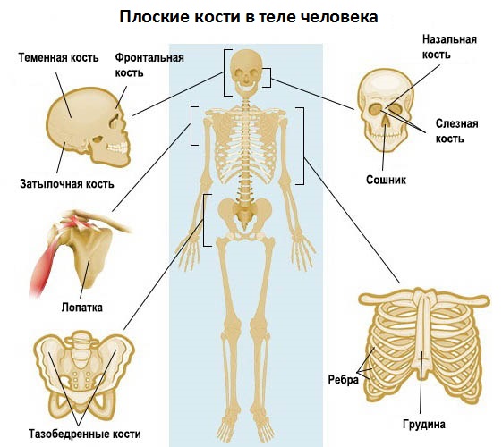
Flat, wide bones provide protection and muscle attachment. These bones are expanded into wide, flat slabs, as in the skull, hip bones, sternum, rib cage and scapula.
The flat bones of the human body are:
- Occipital
- Parietal
- Frontal
- Nasal
- Tearful
- Opener
- Shoulders
- Femoral
- Sternum
- Ribs
Skull and facial bones
The bones of the skull include the occipital bone, two parietal bones, the frontal bone, two temporal bones, sphenoid bone and ethmoid bone. The upper part and both sides of the head are formed by paired parietal bones. The frontal bone forms the forehead, while the occipital bone forms the back of the head. All these thin, curved plates protect the brain in the event of traumatic injury. There are fourteen facial bones, including the jaws, cheekbones, lacrimal, nasal, inferior turbinates, palatines, vomer and lower jaw. Of these, the nasal bones (two oblong shaped bones that form the bridge of the nose), the lacrimal bone (a small bone of the skull that is located in the anterior part of the medial wall of the orbit) and the vomer (a quadrangular-shaped bone that forms the lower and posterior part of the nasal septum) are classified as categories of flat bones.
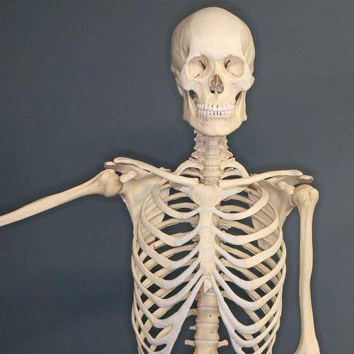
Ribs
The human rib cage consists of twelve pairs of curved, flat bones called ribs, twelve thoracic vertebrae, and a T-shaped bone called the sternum. Ribs are classified into true ribs, false ribs, and floating ribs. The first seven pairs of ribs are called true ribs. The ends of these ribs are attached to the sternum by costal cartilage, which is embedded in connective tissue. The next three pairs of ribs, called false ribs, connect to the costal cartilages with the lowest pair of ribs. The last two pairs of ribs are called floating ribs. They are attached only to the spine and do not connect to the sternum.
Spatula
The scapula is a triangular bone that forms the back of the shoulder girdle. She joins humerus(upper arm bone) in the collarbone. These are flat, paired bones with a large surface area for muscle attachment. The scapula has three angles (lateral, superior, and inferior), three borders (superior, lateral, and medial), three processes (acromion, vertebral column, and coracoid), and two surfaces (costal and posterior).
Sternum
The sternum is a flat T-shaped bone that is located in the upper middle region in the anterior section chest. It is part of the chest. It is attached to the cartilage of the true ribs (the first seven pairs) and the clavicle on both sides. It is convex-shaped in the front and slightly concave in the back.
Femurs
The right and left thigh bones, sacrum and coccyx form the pelvis in the human body. The right and left femurs meet in front at the symphysis pubis, and connect to the sacrum behind. Each pelvic bone consists of 3 parts, which are called the ilium, ischium and pubis. These three bones make up the anterolateral portion of the pelvis. Ilium is the largest of these bones and forms the main section of the hip bone. The ischium forms the lower section of the back, and the pubis forms the lower section in front. These bones are separated in childhood but fuse together in hip joint at the age of 25 years.
Flat bones are important because they not only protect vital organs and tissues, but also provide a large surface area for the attachment of ligaments and tendons. In addition, cancellous bone tissue, which lies between layers of tough, compact bone tissue, also contains red bone marrow.
Skeleton, which is the axial organ musculoskeletal system, contains different types of bones. They differ in shape, structure and functions.
Features of the structure of bone tissue
This type of connective tissue has a typical structure. It consists of cells called osteocytes and a substance that fills the space between them. This fabric is unique in its own way chemical composition and properties. Organic substances, namely collagen fibers, give it elasticity. And mineral salts are strength. For example, femur can withstand loads of several tons. And if inorganic substances are removed from bone tissue, it will easily crumble.
Types of human bones: classification signs
It is based on several signs. These are shape, size and structure. The types of bones also determine the function they will subsequently perform. The shapes are long, short and wide. The former contain a cavity inside filled with yellow bone marrow. This structure provides long strength and lightness. At their ends there is a spongy substance, between the elements of which there is red bone marrow. This is the basis of the body's hematopoietic cells. Short and completely formed by spongy substance.
Bones are also paired and have no analogues in the body. The first group mainly forms the skull. These include the temporal, zygomatic, parietal. Some bones of the belts and free limbs are also paired. These are the clavicles, shoulder blades, radial, humeral, pelvic. Examples of unpaired bones are the frontal, occipital, and mandibular.
Based on their location in the body, the bones of the skeleton and the torso are distinguished. TO last group includes the spine, sternum and ribs. Also, according to this feature, the bones of the belts and free upper and lower extremities are distinguished. There are more than 200 of them in the human body.
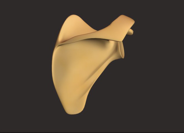
Table: bone types
| Types of bones | Examples | Structural features |
| Long (tubular) | Femoral, tibial, humeral, radial, ulnar | The length of the bones of this species significantly exceeds the width. On top there is a connective tissue layer - the periosteum. Due to it, growth in thickness occurs. At the ends of the bone there is a spongy substance that contains red bone marrow. This is where blood cells are formed. The bone cavity is filled with yellow bone marrow. |
| Short | Frontal and parietal bones of the skull | The length and width of bones of this type are approximately the same. They are entirely formed by a spongy substance that covers a layer of compact substance. |
| Wide (flat) | Sternum, ribs, shoulder blades | The area of the bones exceeds the thickness. They are formed by two plates consisting of a compact substance, between which there is a spongy substance. Due to their large plane, they provide a basis for muscle attachment. |
What are mixed bones?
Very often, due to the complex structure of the bone, it cannot be classified as the main type. They are called mixed. These structures include the vertebrae, sacrum, and clavicle bones. They consist of several parts. Thus, the vertebra is formed by a body and processes, and the main function of this structure is to protect the spinal cord.
Types of bones and features of their connection
All bones in the human body are combined into complex system using connections various types. The way they are attached to each other determines the function of the resulting structure. For example, the flat and wide bones of the skull are connected motionlessly. This method is called a seam. This connection allows you to create reliable protection for the brain. Various types Skeletal bones differ in their characteristics. For example, ulnar, radial and knee joint They move great. This movable connection provides the main function of this structure. It consists in ensuring the movement of individual parts and the skeleton as a whole. The spine, which is the axial structure of the body, is a semi-movable joint. The thing is that between its individual elements there are cartilaginous layers. This structure provides shock absorption when moving.
So, the main types of skeletal bones in the human body are tubular, short and wide. Their main differences lie in the features of the internal structure, shape, type of connection and function performed.
Tubular bones They are long and short and perform the functions of support, protection and movement. Tubular bones have a body, a diaphysis, in the form of a bone tube, the cavity of which is filled in adults with yellow bone marrow. The ends of the tubular bones are called epiphyses. The cells of spongy tissue contain red bone marrow. Between the diaphysis and epiphyses are the metaphyses, which are areas of bone growth in length.
Spongy bones distinguish between long (ribs and sternum) and short (vertebrae, carpal bones, tarsus).
They are constructed of a spongy substance covered with a thin layer of compact. Spongy bones include sesamoid bones (patella, pisiform bone, sesamoid bones of the fingers and toes). They develop in muscle tendons and are auxiliary devices for their work.
Flat bones , forming the roof of the skull, built from two thin plates of a compact substance, between which there is a spongy substance, diploe, containing cavities for veins; the flat bones of the belts are built of spongy substance (scapula, pelvic bones). Flat bones serve as support and protection,
Mixed dice merge from several parts that have different functions, structure and development (bones of the base of the skull, collarbone).
Question 2. Types of bone joints.
All bone connections can be divided into 2 groups:
continuous connections - synarthrosis (immobile or sedentary);
discontinuous joints - diarthrosis or joints (mobile according to function).
The transitional form of bone joints from continuous to discontinuous is characterized by the presence of a small gap, but the absence of an articular capsule, as a result of which this form is called a semi-joint or symphysis.
Continuous connections are synarthrosis.
There are 3 types of synarthrosis:
Syndesmosis is the joining of bones using ligaments (ligaments, membranes, sutures). Example: skull bones.
Synchondrosis is a connection of bones using cartilage tissue (temporary and permanent). The cartilage tissue located between the bones acts as a buffer, softening shocks and shocks. Example: vertebrae, first rib and vertebra.
Synostosis is the joining of bones through bone tissue. Example: pelvic bones.
Discontinuous joints, joints – diarthrosis . At least two are involved in the formation of joints articular surfaces , between which is formed cavity , closed joint capsule . Articular cartilage , covering the articular surfaces of the bones are smooth and elastic, which reduces friction and softens shocks. The articular surfaces correspond or do not correspond to each other. The articular surface of one bone is convex and is the articular head, and the surface of the other bone is correspondingly concave, forming the articular cavity.
The joint capsule is attached to the bones that form the joint. Hermetically closes the joint cavity. It consists of two membranes: outer fibrous and inner synovial. The latter secretes a clear liquid into the joint cavity - synovia, which moisturizes and lubricates the articular surfaces, reducing friction between them. In some joints, the synovial membrane forms, protruding into the joint cavity and containing a significant amount of fat.
Sometimes protrusions or inversions of the synovial membrane are formed - synovial bursae lying near the joint, at the junction of tendons or muscles. Synovial bursae contain synovial fluid and reduce friction between tendons and muscles during movement.
The articular cavity is a hermetically sealed slit-like space between the articular surfaces. Synovial fluid creates a pressure in the joint below atmospheric pressure, which prevents the divergence of the articular surfaces. In addition, synovia is involved in fluid exchange and strengthening of the joint.
The skeleton is divided into the following parts: the skeleton of the body (vertebrae, ribs, sternum), the skeleton of the head (bones of the skull and face), the bones of the limb girdles - upper (scapula, collarbone) and lower (pelvic) and the bones of the free limbs - upper (shoulder, bones forearm and hand) and lower (thigh, leg bones and foot).
The number of individual bones that make up the skeleton of an adult is more than 200, of which 36 - 40 are located along the midline of the body and are unpaired, the rest are paired bones.
Based on their external shape, bones are distinguished into long, short, flat and mixed. However, such a division, established back in the time of Galen, based only on one characteristic (external form) turns out to be one-sided and serves as an example of the formalism of the old descriptive anatomy, as a result of which bones that are completely heterogeneous in their structure, function and origin fall into one group. Thus, the group of flat bones includes the parietal bone, which is a typical integumentary bone that ossifies endesmally, and the scapula, which serves for support and movement, ossifies on the basis of cartilage and is built from ordinary spongy substance.
Pathological processes also occur completely differently in the phalanges and bones of the wrist, although both belong to short bones, or in the femur and rib, which are included in the same group of long bones. Therefore, it is more correct to distinguish bones on the basis of 3 principles on which any anatomical classification should be built: form (structure), function and development.
From this point of view, the following classification of bones can be outlined (M. G. Gain):
- I. Tubular bones
- 1. Long
- 2. Short
II. Spongy bones - 1. Long
- 2. Short
- 3. Sesamoids
III. Flat bones - 1. Skull bones
- 2. Belt bones
IV. Mixed dice
I. Tubular bones. Constructed of a spongy and compact substance that forms a tube with the medullary cavity; perform all 3 functions of the skeleton (support, protection and movement). Of these long tubular bones(shoulder and bones of the forearm, femur and bones of the leg) are struts and long levers of movement and, in addition to the diaphysis, have endochondral foci of ossification in both epiphyses (biepiphyseal bones); short tubular bones(carpal bones, metatarsals, phalanges) represent short levers of movement; Of the epiphyses, the endochondral focus of ossification is present only in one (true) epiphysis (monoepiphyseal bones).
II. Spongy bones. Constructed primarily of a spongy substance covered with a thin layer of compact. Among them there are long spongy bones(ribs and sternum) and short(vertebrae, carpal bones, tarsus). Spongy bones include sesamoid bones, i.e., sesame plants similar to sesame grains, which is where their name comes from (patella, pisiform bone, sesamoid bones of the fingers and toes); their function is auxiliary devices for muscle work; development is endochondral in the thickness of the tendons. Sesamoid bones are located near the joints, participating in their formation and facilitating movements in them, but are not directly connected to the bones of the skeleton.
III. Flat bones:
A) flat bones of the skull(frontal and parietal) perform a predominantly protective function. They are built from 2 thin plates of a compact substance, between which there is diploe, diploе, - a spongy substance containing channels for veins. These bones develop on the basis of connective tissue (integumentary bones);
b) flat bones belts(scapula, pelvic bones) perform the functions of support and protection, built mainly from spongy substance; develop on the basis of cartilage tissue.
-
 April 17, 2015"Marble" cupcake: recipes and cooking methods
 -
 April 17, 2015Recipe without sterilization with onion sautéing
 -
 April 17, 2015Lazy achma in a slow cooker
 -
 April 17, 2015Achma from lavash in a slow cooker


