Number of vertebrae in animals. Spinal column. Slow animals have longer necks
SKELETON
The animal's skeleton consists of axial and peripheral parts. The axial skeleton includes the skeleton of the head (skull) and the skeleton of the stem part of the body. The peripheral skeleton is formed by the bones of the limbs and is divided into the skeleton of the girdles and free limbs.
Lesson 16. SKELETON OF THE BODY TRUNK
Purpose of the lesson: to study the structure of the bones of the stem skeleton.
Only the articulated cranial and caudal processes of the vertebral arches, which act as planar joints, are connected by true articulations. The bony base of the horse's neck area. Ligaments of the neck of the equestrian region. The vertebral ligaments are generally divided into short ones, which unite only the long ones and adjacent vertebrae, which connect the spinal column into a functional unit. The interlinear ligaments extend between the spinous processes and are elastic in the cranial region of the horse. These ligaments prevent dorsal displacement of the vertebral bodies and limit flexion of the spine.
Materials and equipment. Anatomical preparations: complete thoracic segment; first, second, typical and last cervical vertebrae; thoracic, lumbar and caudal vertebrae, sacrum, rib, sternum of cattle, horses, pigs.
The stem skeleton is formed by the bones of the neck, torso and tail. It consists of bony segments that are fully developed in the anterior part of the thoracic region - each segment consists of one vertebra, two ribs and a segment sternum. Reduction of parts of the bone segments occurs both in the cranial and caudal directions. First, the sternum disappears, the ribs are reduced, the remains of which grow to the vertebrae, and then the vertebrae themselves. All vertebrae of the stem skeleton form spinal column(spine), inside it, that is, in spinal canal, the spinal cord is located.
Intertransitional ligaments are among the poorly developed transverse processes in the cervical vertebrae. The long ligaments are classified as: the dorsal longitudinal ligament, which runs in the spinal canal from the odontoid processes of the axis to the sacral bone, on the dorsal portion of the vertebral bodies and adheres to the ligamentous ridges and internal discs. vertebra. The neck ligament is always tense due to the weight of the head and thus reduces the load on the muscles of the head and neck. In the horse, this ligament has its origin in the occipital scale, caudally it transforms into the supraspinatus ligament.
Thorax skeleton cattle consists of 13-14, horses - 17-19, pigs - 14-17 vertebrae, the same number of pairs of ribs and sternum. The diaphragmatic vertebra, through which the center of gravity of the animal passes, is the 13th in cattle, the 15th in horses, and the 11th in pigs.
Thoracic vertebra- vertebra thoracica - cattle (Fig. 36, A) consists of body 8 And arches (arms) 3, which are connected to each other arch roots. Between the body and the arch is the vertebral foramen. On the body, a convex shape is distinguished from the cranial side head 9, from caudal - concave hole 6. The head and fossa of the bodies of two adjacent vertebrae are connected
In the horse, the neck ligament is formed by the neck fluculus and the neck plate, both for development. The nuchal cord communicates the external occipital protuberance, after ligation with the nuchal plate, at the height of the third cervical vertebra, with the spinous processes of the fourth thoracic vertebra into which it is inserted. In the area of the cross, the nuchal ligament becomes wide, forming the cap of the cross. Under the occipital plate, between the cap of the cross and the spinous process of the second and third thoracic vertebrae, there is a supraspinal subligation sac.
Also in the cervical vertebrae, a subligation pocket of the cranial neck of the atlas and a sublimation pocket of the caudal axis gap have been described. The muscle base of the horse's neck area. Muscular, subcutaneous and histological basis of the horse's neck area. Myology of the neck of the Equine region. Under the skin, the surface of the head and trunk is surrounded by fascia, which allows the origin and insertion of muscles and contributes to their ability to glide over it. These fascia are involved as wrapping the organs, in addition to the vessels and nerves in the depths. In this step we will emphasize the superficial fascia of the neck and the deep fascia of the neck.
Rice. 36. Thoracic vertebra:
A- cattle; B- horses; IN- pigs
among themselves. Neutral on the vertebral body there is a weakly expressed ventral ridge 7. On the sides of the head and fossa, smoothed depressions are noticeable - cranial costal fossae 10 And caudal costal fossa 5. Two fossae of adjacent vertebrae form deep costal fossa for connection to the rib head. On the arch there are 13 cranial and caudal articular processes 2, which articulate the arches of two adjacent vertebrae. At the roots of the arches in front, paired cranial vertebral notches 11, and behind - lateral(lateral) vertebrates(intervertebral) holes 12 through which spinal nerves and arteries pass. Transverse processes extending from the arch to the lateral sides, carrying transverse costal fossae(fossae of the transverse processes) 4 And mastoid processes 14. In the dorsal direction it extends from the arch spinous process 1, long at the thoracic vertebrae, directed somewhat backward in the withers area. In the diaphragmatic - 13th vertebra, the spinous process is directed straight upward.
The superficial fascia of the neck can be divided into a superficial and a deep sheet. The superficial sheet covers the superficial muscles of the neck, the ventral muscle of the husk of the neck, the muscle of the spleen, and the insertions into the ligament of the neck. The deep fascia of the neck consists of two leaves, the superficial leaf, which has its origin in the atlas wing, in the longus capitis muscle and in the scalp muscles. It continues ventrally and surrounds the esophagus, the recurrent laryngeal nerve, the vagosympathetic chest, and the common carotid artery. This fascia is inserted cranially into the hyoid bone and into the fascia of the pharynx, caudally into the first pair of ribs and into the sternum.
At the horse B Compared to cattle, the thoracic vertebra has a shorter body, the head and fossa are more flattened, and the ventral ridge is better expressed, resulting in the body taking on a triangular shape. The cranial and caudal costal fossae are deeper, the apices of the spinous processes are thickened and sometimes bifurcated, instead of a paired lateral vertebral foramen there is a paired caudal vertebral notch, which is with the cranial.
The deep leaf is born in the intertransitional muscles and surrounds the longus colli muscle and the longus capitis muscle. The cutaneous muscles, together with the superficial and deep fascia, form an enveloping surface of contractile tension, which has a special functional significance. The superficial sphincter muscle of the neck is located as a narrow transverse muscle strip that passes ventral to the larynx through the neck in a caudal direction. The deep sphincter muscle of the neck extends below the platysma or cutaneous muscle of the face on the side of the neck and head.
the vertebral notch of the anterior vertebra forms intervertebral foramen.
At the pig's IN in comparison with cattle, the body of the thoracic vertebra is more cylindrical in shape, the ventral ridge is not developed, and there is a cross hole 15.
Rib - costa. Cattle have 13-14, horses - 17-19, pigs - 14-16 pairs of ribs. They form the lateral wall of the chest. Cattle rib (see Fig. 37, A) consists of rib bone And costal cartilage. The rib bone has two ends: the dorsal one, facing the vertebra ( vertebral), and ventral, facing the sternum ( sternal). At the vertebral end there are head 1 with articular surfaces for articulation with the costal fossae on the vertebral bodies, neck 2, tubercle 3 with an articular surface for articulation with the transverse costal fossae of the transverse processes, edge angle 4. Rib body 6 wide and flat, convex on the lateral side and concave on the medial side. On the cranial concave edge there is muscle groove 5, on the caudal convex edge - vascular groove 7.
This cutaneous muscle tightens the superficial fascia in the area of the larynx. The cutaneous muscles of the neck are referred to as position and function. They are innervated by the neck branch of the facial nerve. The cutaneous muscles of the neck consist of the superficial sphincter neck muscle, the platysma, the deep sphincter neck muscle and the cutaneous neck muscle, which is a muscular plate on the ventral side of the neck that arises from the sternum and it covers the jugular groove. The neck muscles are located in the back of the neck and in the lateral surface of the neck, some of them are associated with the hyoid apparatus.
Cattle have 8 pairs of sternal (true) ribs, the cartilaginous ends of which articulate with the sternum. The remaining ribs are asternal (false), with their cartilages attached to the cartilages of the ribs in front. On the 2nd-10th ribs, joints form between the costal bone and cartilage.
Horse ribs B compared to cattle they have a more rounded body 6, convex to the lateral side in the form of an arc, edge angle 4 poorly expressed. There are 8 pairs of sternal ribs.
Irrigation of the Cuello del Caballo region. Irrigation of the structures of the head and neck is provided by the branches of blood vessels that pass through the neck, which consist of: the common carotid artery, the external jugular vein, the artery and vertebral vein, the artery and deep cervix, the venous plexus, the vertebral and ventral spinal artery.
Lymph nodes of the horse's neck region. IN cervical spine the horse is described as a superficial cervical lymph node. Innervation of the neck of the Equine region. The spinal nerves contain motor fibers, sensory and autonomic, and at the cervical level it has 8 segments, which are named after the intervertebral canal through which they emerge. At the level of the cervix, two plexuses of cervical nerves have been described: the superior cervical ganglion, the vagus nerve, and the spinal nerve. Areas of cutaneous innervation of the neck, in general, predominate in the neck, metamuscular or segmental organization, which guides its development and morphogenesis.
On the ribs of a pig IN better expressed costal angles 4, as a result of which the bone rib has the appearance of a comma. There are 6-8 pairs of sternal ribs.
The sternum - sternum - forms the lower wall of the chest. In cattle it consists of the manubrium, body and xiphoid process (Fig. 38, A). Handle 1 directed cranially,
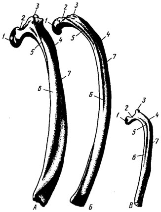
Rice. 37. Rib:
A- cattle; B- horses; IN- pigs
The dermotomes of the first cervical segments are mainly stretched and drawn towards the head, and with them the cutaneous branches of the corresponding nerves, therefore the first two cervical nerves contribute to the innervation of the skin of the occipital region and the back of the ear with the help of the cutaneous endings of the ear with the cutaneous endings of the cervical dorsal branches, with one on the hand, and with the magnetic and transverse rough nerves of the neck, on the other, affecting the skin of the lateral base of the auricle and the venous and intermaxillary regions. The cutaneous region of the large atrial and transverse nerves of the neck completely occupies the areas of the retrouricular, parotid and laryngeal region and in the greater or to a lesser extent overflows their caudal limits.

Rice. 38. Sternum:
A- cattle; B- horses; IN- pigs
has paired pits on the sides for the first costal cartilages. It is articulated with the body by a joint. Body 2 flattened in the dorsoventral direction, widens caudally, consists of six segments connected by cartilage, it has 6 pairs rib tenderloins 3 for articulation with sternal ribs. Xiphoid process 4 directed caudally, xiphoid cartilage 5 in the form of a wide thin plate.
The telltale nerves constitute the terminal division of the ventral branch of the 2nd cervical nerve. Almost the dorsal region of the neck, as well as half of the dorsal collapse of the lateral region, are occupied on the surface of the skin region of the dorsal branches of the cervical nerves. The dermal fillets of these rami, which appear very close to the dorsal midline and more transversely on the horse, present a single caudo-ventral trajectory, especially those with caudal levels, which also appear to be more elongated over a larger area of the corresponding dermotomes.
Horse sternum B has cartilage additives on the handle - falcon 6, which descends to the ventral side of the body, strongly compressed from the sides, in the form sternal crest 7. There are 7 costal notches on the body.
Pig belly IN has a rectangular handle 1 with a common rib notch for the first pair of ribs. On the body of the sternum, consisting of four segments, there are 5 pairs of costal notches. The xiphoid cartilage is short, elongated oval in shape.
For similar reasons for stretching, the first thoracic dermotome and its nerve are topographically included in the dorsal base of the horse's neck. The area in question predominantly affects the skin of the brachycephalic, sternocephalic and tracheal regions. Caudo-dorsal and caudo-ventral shunts of the ventral rami of the 6th cervical nerve, the skin emerges from the caudo-ventral borders of the neck as the supraclavicular nerves are simply or staggered in the dorsal, intermediate and ventral supraclavicular nerves.
Thus, these neural pathways will be the determinants of the supraclavicular nerves that influence the caudal half of the speculum region and, to a greater or lesser extent, the caudal sulcus of the lateral and ventral cervical regions. The dissection that shows these links allows the establishment of precise anatomical planes in two large areas or substrates in which, as a rule, we divide the horse's neck into its topographic analysis, that is, into the dorsolateral substrate and the ventral substrate. The demarcation will be given by a line projected by a series of tangible cervical transverse processes.
Lumbar skeleton in cattle it consists of 6, in a horse - from 5-6, in a pig - from 5-7 lumbar vertebrae.
Lumbar vertebra- vertebra lumbalis - cattle (Fig. 39, A) has a long body 1 with flat head And hole 2. Well expressed ventral ridge 3, there is a deep caudal vertebral notch 4, sometimes forming the lateral (side) vertebral foramen. Between the body and the arch - vertebral foramen 9. There is a low
Skin and subcutaneous tissue of the cervical region del Caballo. The neck is the surface covered with skin, the subcutaneous layer on the dorsal border, the fatty crest of the occiput. The neck or crest of the Cresti is rounded and only the upper part of the spinous processes is palpable or may be covered with fat. Neck circumference may represent fat accumulation around the lower region. Neck perimeter measurements are important in assessing the horse's body condition.
Anatomical regions of the horse's neck. Three broad dorsal, lateral and ventral regions of the cervix are generally described, with three other regions described, of which two correspond to the cephalic limb and the third to the base of the neck; however, from a practical point of view, it is often referred to more specific regions, such as the brachiocephalic, sternocephalic, retroarthrial, pharyngeal, and tracheal regions.
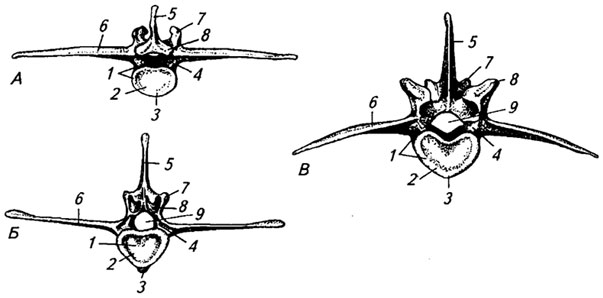
Rice. 39. Lumbar vertebra:
A- cattle; B- horses; IN- pigs
spinous process 5, long transverse costal processes 6, formed as a result of fusion of the transverse process with a reduced rib, there are articular processes; of which cranial 7 semi-arc shape, caudal 8- cylindrical.
Anatomical areas of the horse's neck. X-ray of the cervix, occipitoatloid and first joints. cervical vertebra. It extends from the back of the head to the spines of the chest and back. In the horse it becomes more and more extensive as it approaches the base of the neck, so that the horse understands here the entire surface, which is designated externally as the “tables of the neck”, also highlighting the dorsal edge of the neck which gives the implantation of the neck bristles or “creaner”, which is determined by the cervical cord. This ligamentous cord and adjacent muscles form the anatomical basis of the region.
At the horse B body 1 lumbar vertebra is shorter, ventral ridge 3 expressed only in the first vertebrae, there are no lateral foramina, the spinous process is higher and narrower. U cranial 7 And caudal articular The articular platforms of the processes are smooth; on the last two or three transverse costal processes there are articular platforms.
This is the most extensive of all areas of the cervix, since the entire surface of the cervical tables belonging to the horse, the dorsal region and brachycephaly are collected here. On a horse, if we deal with the jugular groove, which contains the jugular vein and the jugular fossa or limited depression of the caudal end of the groove. The brachycephalic region contains most of the lateral region that touches us.
In fact, they represent three areas which, from the point of view of application, deserve to be described as more or less visible areas in the form we are studying. The latter limits rostrally to the subhydoid region of the intermaxillary space.
At the pig's IN the ends of the transverse costal processes are directed downwards, at their base there are either lateral openings, either notches or fossae, the vertebral heads are flat.
Skeleton of the sacrum formed by sacral vertebrae fused into one bone.
Sacral bone - os sacrum - of cattle (Fig. 40, A) consists of five fused vertebrae. Their bodies formed body of sacral bone 1, on which the sutures (transverse lines) from the fusion of the vertebrae are visible. The body narrows caudally and curves dorsally. The vertebral foramina are combined into sacral canal 9. Instead of cranial and caudal vertebral notches, dorsal 2 And ventral sacral foramina. The arches and spinous processes also fused, forming the median sacral crest 4. The transverse costal processes of the first two sacral vertebrae formed wings of the sacrum 5, compressed from front to back and having a rough ear-shaped surface 6 for articulation with the ilium
Ventral subgroup Retrouricular and parotid. Both regions continue from the caudal base of the ear to the region of the larynx, which we have left inscribed in the ventral region of the neck. The first corresponds to the small territory that remains between the indicated atrial base and the wing of the atlas, so its anatomical base mainly belongs to the oblique cranial muscle. Ventrally, it expands the parotid region, which has its anatomical basis in the salivary gland and a very precise supporting cranial limit: the caudal border of the ramus.
It is to this limit that depression can be visible in the region either with the naked eye or through palpation, this is the retro-mandibular fossa, which in research practice projects the pharyngeal region in relation to the intermaxillary space. Thus, we call the parotid, pharyngeal and laryngeal topography together the “cervicofacial transit territory”, of particular anatomical and surgical significance.
pelvic girdle. The transverse processes of the remaining vertebrae were reduced and formed side parts 3 sacral bone. On the first sacral vertebra on the sides of heads 8 preserved cranial articular processes 10 crescent-shaped, on the last - caudal articular processes in the shape of a cylinder, under the head is cape 7.
At the horse B The sacrum bone consists of 5-6 fused vertebrae. Body 1 its direct, wings 5 are located in the frontal plane, the ends are slightly directed forward, the spinous processes are higher than those of cattle, their ends are expanded, and sometimes bifurcated and do not grow together. On the wings, in addition to the auricular, there is an anterior articular surface 11 for articulation with the same surface of the last lumbar vertebra. The cranial articular processes are straight.
At the pig's IN The sacrum bone consists of 4 fused vertebrae. The spinous processes have been reduced; between the arches of the fused vertebrae there are interarch holes 12, the wings are directed in the sagittal plane.
Tail skeleton formed by the caudal vertebrae - vertebrae caudales. Cattle have 18-21 of them. The vertebral bodies are long, rudiments of arches are visible on the first 3-5 vertebrae, and on the ventral surface of the body - hemal arches for the passage of the caudal artery, which then pass into hemal processes, visible up to the 10th vertebra. The transverse processes are short, wide, and curved ventrally. Towards the end of the tail the vertebrae are greatly reduced.

Rice. 40. Sacrum bone:
A- cattle; B- horses; IN- pigs
A horse has 15-20 caudal vertebrae. The vertebral body is short, massive, its width is almost equal to its length, the rudiment of the arch is weakly expressed and gradually disappears completely, there are no hemal arches.
The pig has 20-23 caudal vertebrae; they are small, the arches are well developed and protrude caudally from the body.
Neck skeleton consists of seven vertebrae. The first and second cervical vertebrae are very different from the rest. The 3rd-5th are typical, the sixth has a changed shape of the transverse costal process, and the seventh has a high spinous process, an unbifurcated transverse costal process and caudal costal fossae for articulation with the first rib, there is no intertransverse foramen.
Typical cervical vertebra cattle (Fig. 41, A) has a relatively short body 4, well expressed head 9 And hole 3, deep cranial 8 And caudal vertebral notches 2. Spinous processes 11 increase from the third to the seventh vertebrae, cranial 10 And caudal articular processes 1 flat, well developed, transverse costal processes 7 bifurcated (one part is directed ventrally, the other dorsally). Between the transverse costal processes and the roots of the arch there is intertransverse(vertebral-costal) hole 6. The ventral ridge is absent.
At the horse B body 4 elongated, head 9 convex, hole concave, spinous processes very weakly expressed, and ventral ridge 5-very much, transverse costal processes bifurcated in the craniocaudal direction.
At the pig's B head 9 And hole 3 flattened. On the transverse costal processes there is cross hole.
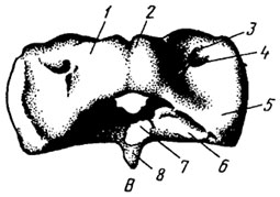
Second cervical vertebra(axial) - axis - cattle (Fig. 42) has body 4, arc and wide vertebral foramen. Instead of the head there is tooth, or odontoid process 9, semi-lunar shape. On both sides of it are
articular surfaces 8 for articulation with the atlas. The vertebral fossa is deep 3 . The spinous process is rectangular in shape and is called crest 1, caudal articular processes 2 isolated, instead of cranial vertebral notches intervertebral foramina 7, transverse costal processes 5 are not forked and have intertransverse holes 6.
At the horse B odontoid process 9 chisel-shaped, its end is pointed, articular surface 8 borders the process from the sides and below, crest 1 arched, bifurcated and bears caudal articular processes 2, ventral ridge 10 highly developed.
At the pig's IN the body of the axial vertebra is short, the odontoid process is cone-shaped, the crest is very high and raised towards the posterior edge.
The first cervical vertebra (atlas) - atlas - of cattle (Fig. 43, A) has the shape of a ring and consists of two arcs: dorsal 1 with dorsal tubercle 2 And ventral 7 With ventral tubercle 8. The body is reduced. At the caudal end of the atlas there is a flattened articular surface 6 for articulation with the odontoid process of the second cervical vertebra. At the cranial end there are ellipsoidal shapes cranial glenoid fossae for articulation with the condyles of the occipital bone.
|
|
|
|
|
Rice. 43. First cervical vertebra: A- cattle; B- horses; IN- pigs |

The transverse costal processes of the atlas are in the form of wide thin plates and are called wings 5. Located on the wings wing holes 4, they pass from the dorsal to the ventral side and are located laterally intervertebral foramen 3.
At the horse B the wings of the atlas are lowered ventrally. They have additional intertransverse (transverse costal) holes 9.
At the pig's IN there is a transverse canal on the wings, the ventral tubercle is strongly developed and projects caudally.
Phylogenetic transformations. In fish, the spine is distinguished only by the trunk and caudal sections. All the vertebrae in it are almost identical. The trunk vertebrae bear ribs.
Already in the first terrestrial vertebrates - amphibians, the hind limbs rest through the pelvic bones on the processes of one of the vertebrae, formed by the accreting sacral ribs. This vertebra is located on the border between the trunk and caudal vertebrae and is called the sacral vertebra. The first vertebra, the only cervical one, forms a movable articulation with the skull.
In reptiles, the true ribs connect to the sternum to form the rib cage, and the trunk spine is divided into the thoracic and lumbar spine. These are followed by the sacral and caudal sections of the spine. The cervical ribs are rudimentary, in the lumbar region they are poorly developed, and in the sacral region, formed by two vertebrae, they fuse with them, giving support to the pelvis.
In mammals and humans, minor remains of ribs in the cervical and lumbar vertebrae are fused with the transverse processes, and in the sacrum they form its lateral parts (Fig. 12). Mammals (with very few exceptions) have seven cervical vertebrae. Due to the sharply increasing importance of the hind limbs in locomotion, the pelvis no longer articulates with two, but with a large number of vertebrae. This enlarges the sacral region.
If the ribs are phylogenetically very ancient and were already present in fish, then the real sternum appeared much later, in connection with the complete transition of vertebrates to life on land.
In amphibians it is still small, but quite developed in reptiles and mammals.
Human spine. The human spine, or spinal column, consists of 33-34 vertebrae (vertebrae), metamerically following each other (Fig. 18). The spine is divided into sections: cervical (7 vertebrae), thoracic (12 vertebrae), lumbar (4-5 vertebrae). The sacral vertebrae fuse into one bone - the sacrum, and the coccygeal vertebrae - into the coccyx. Therefore, in an adult, the spine consists of 24 separate vertebrae, the sacrum and the coccyx.
The spine is the main core of the body and its support. It protects the spinal cord, forms part of the walls of the chest, abdominal and pelvic cavities and, finally, is involved in the movement of the torso and head. The structure of the vertebrae corresponds to these functions.
Vertebra has a massive supporting part - body, arc, which consists of two symmetrical halves, closing the vertebral foramen together with the body, and extending from the arch shoots(Atl., 3, D and 4, D). Some processes serve as attachment sites for muscles - unpaired spinous process, rear-facing, and paired transverse processes, directed to the sides; others articulate with adjacent vertebrae - paired superior and inferior articular processes. The vertebral foramina together form spinal canal, which houses the spinal cord. On the arches, at the point of their transition into the vertebral body, there are intervertebral notches above and below (the lower one is deeper). The notches of adjacent vertebrae form intervertebral foramen, through which spinal nerves and vessels pass.
Along with the general characteristics, the vertebrae of different parts of the spine have differences, most sharply noticeable in the middle parts of each part.
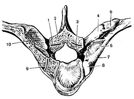
Thoracic vertebrae are the most typical, since only in the thoracic region the segmental structure is completely preserved. They differ from other vertebrae in their articular costal semi-pits(on the I, XI and XII vertebrae - pits), which are located on the lateral surfaces of their body, above and below the base of the arch (Atl., 3, D). Two semi-fossae of adjacent vertebrae form a fossa that articulates with the head of the rib. At the end of the transverse processes of the first ten vertebrae there is articular surfaces, with which the tubercles of the ribs articulate. The spinous processes are directed downward and overlap each other, which is especially pronounced in the middle four thoracic vertebrae. This makes the thoracic spine less mobile. The weight of the vertebral body gradually increases towards the lumbar region (Fig. 13).
Cervical vertebrae preserved minor rudiments from the ribs, fused with transverse processes, which are therefore called transverse costal(Atl., 3, G). The base of the latter has a hole. The part of the process that borders the opening in front is the remainder of the rib. Transverse costal openings all cervical vertebrae form an intermittent canal. It serves to protect the vertebral artery, which passes through it to the brain, and the vein of the same name. The bodies of the cervical vertebrae are less massive than the bodies of the thoracic vertebrae, and their upper and lower surfaces are saddle-shaped, which causes significant mobility of the neck. The vertebral foramina are large, the arches are thin. The spinous processes (with the exception of the process of the VII vertebra) are shorter than in the thoracic region and bifurcated at the end, which increases the area of attachment of numerous muscles to them. The first two cervical vertebrae are sharply separated from the rest.
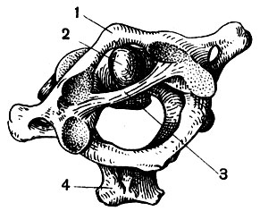
Atlant- the first cervical vertebra - has the shape of a ring (Atl., 3, A, B). The place of the body is occupied anterior arc, on its convex part is located anterior tubercle. On the side facing the inside of the wide vertebral foramen, the articular fossa for the odontoid process of the second vertebra is noticeable. On posterior arch, corresponding to the arches of other vertebrae, only a weak protrusion has been preserved from the spinous process - posterior tubercle. Instead of the superior articular processes, oval superior ones are located on the side glenoid fossae, which articulate with the condyles of the occipital bone. The role of the lower articular processes is played by the fossae that articulate with the second vertebra.
Epistrophy, or axial vertebra, differs from typical cervical vertebrae in the development of a process on the upper part of the body - tooth, around which the atlas rotates along with the skull (Atl., 3, B). This process arises during the uterine period of development by accretion to the epistrophy of most of the body of the atlas. Instead of the superior articular processes, there are slightly convex articular surfaces on the sides of the odontoid process.
Lumbar vertebrae, especially the latter, massive and distinguished by elongated sides transverse costal processes(Atl., 4, D) - the product of the fusion of the transverse processes and rudiments of the lumbar ribs. Small processes on the arch and superior articular processes increase the area of attachment of the powerful back muscles.
Sacrum(sacrum) is shaped like a triangle, with the base pointing upward and the apex pointing downward (Atl., 4, A, B). On the anterior surface of the sacrum, smoothed, concave and facing the pelvic cavity, there are four transverse rough lines- traces of fusion of the sacral vertebral bodies. Four pairs open here anterior sacral foramina. On the back, convex surface there are lumpy median sacral ridge(merged spinous processes), two parallel to it articular crest(merged articular processes), and lateral to them - lateral ridges(merged transverse processes). Four pairs open between the articular and lateral ridges posterior sacral foramina. Located outside the lateral ridges, the lateral parts of the sacrum (fused rudiments of the sacral ribs) articulate with pelvic bones through ear-shaped surfaces. Sharply tapering at the bottom sacral canal serves as a continuation of the spinal canal.
The sacrum of men is longer, narrower and more curved than that of women.
Coccyx(coccygenm) consists of 4 (less often 3 or 5) rudimentary vertebrae fused in an adult, preserving only the body (Atl., 4, C, D). It corresponds to the skeleton of the tail of vertebrates. The tip has the shape of a pyramid, its base facing the sacrum, on which the underdeveloped upper articular and transverse processes of the first vertebra protrude.
Rib cage consists of the sternum and ribs, which are connected to the spinal column at the back.
Ribs(costae) among 12 bunks have the appearance of narrow bone plates, strongly curved and somewhat flattened. Their lower edge is pointed (Fig. 14). The posterior end of each rib articulates with thoracic vertebra with the help heads and tubercles, separated from each other by a narrowed part - neck. The last two ribs (XI and XII) lack tubercles. The first rib is located almost in a horizontal plane (Fig. 15), sharply curved, and a slight elevation protrudes on its upper surface - scalene tubercle(named after the muscle attached here). The anterior ends of the ribs are cartilaginous. Cartilages of the I-VII pairs of ribs ( true) articulate with the sternum. VIII and IX bunks ( false ribs) are connected by their cartilages to the cartilage of the overlying rib, forming costal arch. The cartilages of the X pair sometimes enter it, but more often, like the cartilages of the XI and XII pairs, they end freely in the abdominal muscles ( oscillating ribs). Rarely (2% of people) there are 13 pairs of ribs. In these cases, only 4 lumbar vertebrae remain, since the first of them turns into the XIII thoracic vertebrae. Very rarely there are 11 pairs of ribs (then there are 6 lumbar vertebrae), as well as cervical ribs (on the last cervical vertebra). In general, in the structure of the outer vertebrae of each section there are structural features that are transitional to the neighboring section.

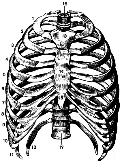
Sternum(sternum) - a flat unpaired bone consisting of an upper part - handles, middle part - body And xiphoid process, which varies greatly in size and shape and often does not live up to its name (Fig. 15). These sections are initially delimited by cartilaginous layers, but with age (after 30 years) they begin to fuse with each other. On the sides of the handle there are notches in which the connection occurs with the collarbones and the first pair of ribs. The upper edge bears an unpaired jugular notch(it can be easily felt through the skin). Along the edges of the body of the sternum, notches are also visible - the junctions with the cartilages of the II-VII pair of ribs.
Women's sternums are usually relatively shorter than men's.
Ontogenesis. During the fourth week of development, a paired chain of sac-like protrusions of mesoderm, called somites, or primary segments. The medial part of each of them forms a sclerotome, and the dorsal and lateral parts - myotome. A membranous axial skeleton develops from the sclerotome, covering the neural tube and notochord (Fig. 65). But in the process of subsequent development, the notochord loses its supporting significance and is preserved only in the form of insignificant remains between the vertebrae, inside the intervertebral discs. At the 5th week of development of the human embryo, the membranous stage passes into cartilage and the connective tissue around the chord and neural tube is replaced by cartilage, which then grows together into rings and form the vertebrae.
The last, bone stage begins from the 3rd month of intrauterine development, when foci of ossification appear in each cartilaginous vertebra - one enchondral in the body and a pair of perichondral in the arch. However, cartilage tissue outpaces bone tissue in growth for a long time and in a newborn accounts for half of the total mass of the spine. The fusion of paired bone foci in the arch, and then the arch with the vertebral body, occurs at the age of 3 to 8 years, in the sacrum - by 10 years. The coccygeal vertebrae ossify only at the age of 14, and the sacral vertebrae fuse with each other between 17 and 25 years. By the age of 10, ring-shaped epiphyses (merge with the vertebra at 22-24 years) and small additional foci of ossification appear along the edges of the upper and lower surfaces of the vertebral bodies.
The ribs and sternum also go through three stages in ontogenesis. The anterior (ventral) ends of the rib cartilages on each side first fuse together. As a result, paired strips appear, which then close together to form the cartilaginous sternum. In the ribs, ossification begins earlier than in the spine, and in the sternum - at recent months intrauterine life. The newborn has a sternum made of cartilage with paired and unpaired bony foci. Later they turn into bone, which consists of several bone segments connected by cartilage, which do not fuse together in adults. After 30 years, the costal cartilages begin to calcify, and in old age they even ossify. This difference in the timing of ossification repeats the phylogenetic sequence in the development of these parts of the skeleton.
Connection of the trunk bones. In the skeleton of the body, the bones are connected through joints and synarthrosis.
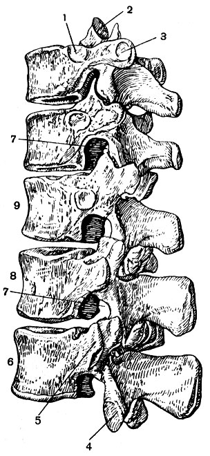
The connection between the vertebral bodies (from the second cervical to the sacrum) is carried out intervertebral discs(Atl., 5, A). Each of them is formed by fibrous cartilage, the bundles of which ring-shapedly surround the elastic nucleus pulposus(remaining chord). The intervertebral discs are firmly fused with the plates of hyaline cartilage covering the top and bottom of the vertebral bodies. Between the vertebral bodies, a kind of semi-joint appears with a nucleus pulposus between the cartilages. The discs make up at least a quarter of the total length above the sacral part of the spine. They are especially thick in the lumbar region. This connection of the vertebrae gives the spine spring-like properties, that is, it softens shocks and at the same time gives it greater flexibility.
Between the articular processes of all vertebrae, real, albeit inactive, articulations are formed. In the cervical and thoracic regions they are classified as flat, and in the lumbar - as cylindrical joints.
Along the anterior surface of the bodies of all vertebrae, starting from the occipital bone and the atlas, stretches anterior longitudinal ligament, and on the back (inside the spinal canal) - posterior longitudinal ligament(Atl., 5, A). Adjacent vertebrae are connected by short intertransverse, interspinous and interarcanal, or yellow, bundles. The elasticity of the ligaments facilitates the work of the muscles that straighten the torso.
Stretches along the spinous processes supraspinous ligament, turning on the neck into a wide nuchal ligament, growing to the occipital bone.
In the sacral and coccygeal sections, the vertebrae are fused using synostoses into complex bones - the sacrum and coccyx.
Atlanto-occipital joint(articulatio atlantooccipitalis) is located between the spine and the skull. This is a paired, combined joint, which is formed by the condyles of the occipital bone and the superior articular fossae of the atlas. It belongs to the ellipsoidal biaxial joints and provides rocking and nodding movements of the head.
The atlas is articulated with the second cervical vertebra by two joints (articiilationes atlantoaxialis). One of them - a pair - is formed by the lower articular fossae of the atlas and the upper surfaces of the axial vertebra. The other joint - unpaired - is formed by the tooth of the axial vertebra and the anterior arch of the atlas, which are firmly pressed to each other by the transverse ligament of the atlas (Fig. 16). The tooth joint of the axial vertebra is classified as uniaxial cylindrical with a vertical axis of rotation. In this joint, the head turns (together with the atlas) to the right and left.
All ribs are connected to the bodies of the thoracic vertebrae by their heads (Fig. 14). The first ten pairs of ribs are also connected to the transverse processes of the vertebrae with the help of tubercles (Fig. 17). Both joints of each rib are combined, that is, movements in them are performed simultaneously. In this case, the necks of the ribs almost do not shift (the axis of rotation passes through them), and the anterior ends of the ribs move forward and to the sides, which increases the volume of the chest. The cartilages of the 1st bunk of the ribs grow together with the sternum, and the II - VII bunks articulate with it with stiff joints (Atl., 6).
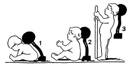
Skeleton of the torso. The connections of the vertebrae (from the second cervical to the sacrum) by intervertebral discs, paired joints and ligaments (Atl., 5) transform the spine into an elastic rod that allows separate or combined movements around the frontal, sagittal and vertical axes (flexion and extension, bending to the side, turning ). Small movements between individual vertebrae, when summed up, provide significant mobility of the spine. The thoracic region is the least mobile due to the presence of ribs, the bevel of the spinous processes and the thinness of the intervertebral discs. The spine makes up about 40% of the total body length and has four curves in the sagittal plane (Fig. 18). Two of them are convexly facing forward ( cervical and lumbar lordosis) and two - back ( thoracic and sacrococcygeal kyphosis). Lordosis and kyphosis balance each other and provide the overall vertical direction of the long axis of the spine. The bends are caused by gravity, muscle tone and a certain wedge shape of the intervertebral discs (in the area of lordosis they are thicker in the front, and in the area of kyphosis - in the back). Lordoses are specific features of the human spine associated with the vertical position of the body. They vary somewhat depending on muscle tone, degree of stomach filling, posture, etc.
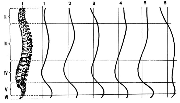
Rice. 18. Spine (left), its functional and age-related curves (lines on the right): I - atlas; II - cervical; III - chest and IV - lumbar region s; V - sacrum; VI - coccyx; 1 - on an empty stomach; 2 - with a full stomach; 3 - with the head down; 4 - with arms extended forward; 5 - at attention young man; 6 - the old man
A newborn baby's spine is almost straight. Cervical lordosis occurs when the child begins to hold his head, that is, actively resist hanging it forward. Later, when the child begins to sit, and then stand and walk, lumbar lordosis appears (Fig. 19), which is finally formed by the age of 15. Intervertebral discs in children are less elastic than in adults, but the spine as a whole is more flexible due to incomplete ossification of the vertebrae. Therefore, when standing, a child spends more muscle effort on fixing the spine than an adult and gets tired faster.
Curvature of the spine to the side - scoliosis, often developing in schoolchildren, is associated with age characteristics vertebral bodies and intervertebral discs (their pliability to deformation) and weakness of the back muscles. Scoliosis occurs when hygiene standards related to the height of desks, strength and uniformity of classroom lighting, individual visual and auditory characteristics of students, etc. are not observed.
Different parts of the spine grow unevenly in length. The lumbar region develops faster than others, and the cervical region develops more slowly.
With old age, the spine shortens (sometimes by 10%) due to a decrease in the height of the vertebral bodies and thinning of the intervertebral discs. Often the curvature of the thoracic region increases significantly and a senile hump appears.
The rib cage forms the bony base of the wall of the chest cavity. The costal cartilages give it elasticity. The rib cage protects the heart, lungs, liver and serves as the attachment point for the respiratory muscles and muscles of the upper limbs.
The shape of the chest is compared to a cone, which has a truncated upper end and an obliquely cut base facing downwards.
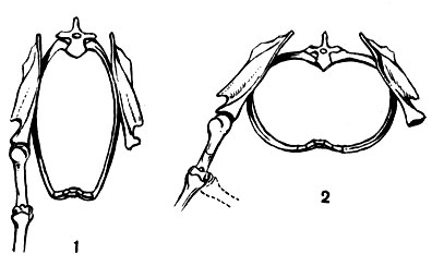
The sagittal size of the chest is always smaller than the transverse size; in a horizontal section it is kidney-shaped (Fig. 20). This shape of the chest is unique to humans and arose in connection with the transformation of the forelimbs of vertebrates into an organ of grasping and then labor. In most animals, the chest is compressed from the sides. Newborns retain traces of similarity with this phylogenetically primary form (Fig. 21, 1). Even schoolchildren junior classes The greater roundness of the chest and less inclination of the ribs are still clearly noticeable than in adults. This is one of the reasons why children breathe less deeply but more frequently. Children with a poorly developed muscular system and weak lungs often have a flattened chest, which appears to be in a collapsed state. For such children, special physical exercises are important. In rickets, the sternum protrudes sharply forward (“chicken breast”). Women's chests are often shorter and more rounded than men's.
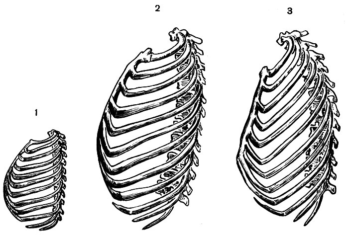
In old people, due to weakening of the muscles that extend the trunk, kyphosis of the thoracic spine increases. The chest shortens and lowers: the anteroposterior size increases (Fig. 21, 3), and the transverse size decreases; The curvature of the ribs becomes less, and they take a more oblique position. All these changes, as well as calcification of the costal cartilages, limit the range of movements of the chest: the difference in its circumference at maximum inhalation and exhalation in old people is 5 cm, and in young people - up to 10 cm.
-
 April 17, 2015"Marble" cupcake: recipes and cooking methods
 -
 April 17, 2015Recipe without sterilization with onion sautéing
 -
 April 17, 2015Lazy achma in a slow cooker
 -
 April 17, 2015Achma from lavash in a slow cooker


