Structure and functions of the bone skeleton. Human skeleton. bone structure
Skeleton(from the Greek skeleton - dried) of a person is a collection of bones connected to each other in a certain way. The adult human skeleton consists of approximately 205 bones. The skeleton (Fig. 12) has three sections: the skeleton of the body, the skeleton of the skull and the skeleton of the limbs (Table 2).
Rice. 12. Human skeleton (front view):
1 - brain skull, 2 - facial skull, 3 - bones of the upper limb girdle, 4 - humerus, 5 - forearm bones, 6 - hand bones, 7 - chest 8 - spinal column, 9 - lower limb girdle bones, 10 - femur , 11 – shin bones, 12 – foot bones
| Bones of body parts | Names of bones and their number |
| Torso bones | Vertebrae – 31 – 33 cervical – 7 thoracic – 12 lumbar – 5 sacrum (5 fused sacral vertebrae) coccyx (3 – 5 coccygeal vertebrae) Ribs – 12 pairs Sternum |
| Skull bones | 23 bones, including unpaired ones - frontal, occipital, sphenoid, lower jaw, hyoid bone and paired bones - parietal, temporal, zygomatic, etc. |
| Bones of the upper limb | 32 bones in one upper limb: clavicle, scapula, humerus radius carpal bones – 8 metacarpal bones – 5 phalanges of fingers – 14 |
| Bones of the lower limb | 31 bones in one lower limb: pelvic bone, femur, patella, tibia, fibula, tarsal bones – 7 metatarsal bones – 5 phalanges – 14 |
The skeleton of the body consists of vertebrae that form the spine and bones chest. Each segment of the human body skeleton is formed by a vertebra, and in the thoracic region also by a pair of ribs and a section of the sternum.
The skeleton of the head, the skull, protects the brain, sensory organs and serves as a support for the initial sections of the digestive and respiratory organs. The skull is conventionally divided into two sections - the brain and the facial.
The skeleton of the upper and lower extremities is divided into the skeleton of the free limb and the skeleton of the belt. Skeleton of the upper limb girdle ( shoulder girdle) consists of two paired bones - the scapula and the clavicle, and the skeleton of the free upper limb - of three sections: the humerus, the bones of the forearm and the bones of the hand.
The skeleton of the lower limb girdle (pelvic girdle) consists of a paired pelvic bone, and the skeleton of the free lower limb is divided into three sections: the femur, the bones of the lower leg and the bones of the foot. Each bone is an independent organ that performs a specific function.
The bones of the skeleton differ in shape and structure. Distinguish tubular, spongy, flat, mixed And air bones(Fig. 13).
Rice. 13. Types of bones:
1 – long (tubular) bone, 2 – flat bone, 3 – spongy (short) bones, 4 – mixed bone
Tubular bones are divided into long (humerus, femur, bones of the forearm and tibia) and short (tarsal and metatarsal bones, phalanges of the fingers). Bones, with the exception of articular surfaces, are covered with a connective tissue membrane - periosteum, which performs bone-forming and protective functions. The periosteum is firmly fused to the bone with the help of connective tissue fibers penetrating deep into the bone. The outer layer of the periosteum is coarsely fibrous, consisting of complexly intertwined fibers and connective tissue cells. This layer contains many blood and lymphatic vessels, nerve fibers that ensure the vital functions of the bone. The inner layer of the periosteum is thin and contains cells from which osteoblasts are formed - young bone cells. Due to the bone-forming function of the periosteum, the bone grows in thickness and heals during fractures.
Inside the bones there are medullary cavity(in tubular bones) and cells of spongy substance, which contains bone marrow. In a newborn and in childhood, the bone marrow cavities are filled with red bone marrow, which performs blood-forming and protective functions. Red bone marrow stem cells form blood cells (erythrocytes, leukocytes) and cells immune system(lymphocytes). In an adult, red bone marrow is stored only in the cells of spongy bone. Other bone cavities contain fatty yellow bone marrow that has lost its function.
On the side of the medullary cavity and cells, the bone is covered with a thin connective tissue plate - endostome, also producing bone tissue.
The bones of the skeleton and the skeleton in the whole body perform supporting, motor and protective functions. Bones are also a depot for minerals - phosphorus, calcium, iron, copper and other trace elements.
Bone Strength ensured by the presence of organic and non-organic organic matter, as well as the structure of bone tissue. In terms of hardness and elasticity, bones can be compared with bronze and cast iron. The compact and spongy substance of bones is built from bone tissue. Compact (dense) bone substance forms the outer layer of each bone. spongy substance, formed by bone crossbars (beams), is located under the compact substance. U tubular bones in the area of their body (diaphysis), the compact bone substance is thick (up to 1 cm). At the ends of tubular bones and flat bones and other bones, this layer is thin. The compact bone substance is penetrated by a system of bone canals in which blood vessels and nerve fibers(Fig. 14).
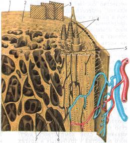
Rice. 14. Scheme of the structure of the tubular bone:
1 - periosteum, 2 - compact bone substance, 3 - layer of outer surrounding plates, 4 - osteons, 5 - layer of internal surrounding plates, 6 - medullary cavity, 7 - bone crossbars of cancellous bone substance.
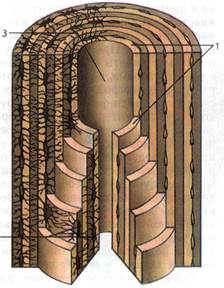 Each bone canal (osteon canal) is surrounded by concentric plates in the form of 4 to 20 thin tubes inserted into one another. The system of such tubes together with the tubule is called osteona, or Haversian system(Fig. 15). The spaces between osteons are occupied by intermediate, or intercalary, plates, which, during bone restructuring due to changing physical load, serve as material for the formation of new osteons. The surface layer of compact bone substance is represented by the outer surrounding plates, which are a product of the bone-forming function of the periosteum.
Rice. 15. Structure of osteon in a section: 1 – osteon plates, 2 – bone cells (osteocytes), 3 – center channel(osteon channel)
The inner layer of bone, bordering the medullary cavity, is formed by internal surrounding plates and covered with fibrous connective tissue - endosteum.
Spongy bone substance located under the compact, is located at the ends of the tubular bones - the epiphyses, in the bodies of spongy, mixed bones, in flat and pneumatic bones. Spongy bone substance consists of bone crossbars intersecting with each other in different directions. Their distribution corresponds to the direction of the main lines of compression (pressure) and tension acting on the bone (Fig. 16).
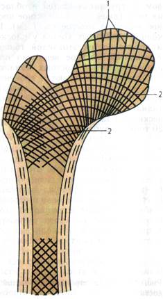 Rice. 16. Scheme of the location of bone crossbars in the spongy substance of bones (cutting of the upper end of the femur): 1 – compression (pressure) lines, 2 – tension lines
This arrangement of the bone crossbars at an angle to each other ensures uniform distribution of pressure and muscle force on the skeletal bones.
Bone is highly plastic. Depending on the load on the bones, the number of osteons increases or decreases, and their location in the compact substance changes. With constant muscle load, sports, and physical labor, the number of osteons and their sizes increase, the layer of compact bone substance in tubular and other bones thickens, and the bone marrow cavities narrow. The bone crossbars (beams) of the spongy substance also thicken and acquire a more complex structure (branch). At the same time, the bones become thicker and stronger. With a decrease in physical (muscle) activity, a sedentary lifestyle, and prolonged bed rest during illness, the bones become thinner and weaker.
Organic and inorganic substances also provide bone strength. Organic substances give bones flexibility and elasticity.
Inorganic substances (calcium phosphate, calcium carbonate and other salts) give bones hardness. In living bone, organic substances account for about 60% of its mass, the rest belongs to inorganic compounds.
The influence of organic and inorganic substances on the strength properties of bones can be tested experimentally. Once the organic matter is removed by roasting the bone over a fire, it becomes brittle. Removing inorganic substances (salts) from the bone by keeping the bone in acid makes the bone soft and flexible. The combination of the hardness of inorganic compounds with the elasticity of organic compounds ensures the strength of bones.
The skeleton is divided into four sections: the skeleton of the body, the skeleton of the head (skull), the skeleton of the upper and lower extremities:
Skeleton of the torso
make up the spinal column and rib cage (12 pairs of ribs and sternum):a) Spinal column
is like the axis of the whole body; it connects to the ribs, to the bones pelvic girdle and with a skull. There are cervical (7 vertebrae), thoracic (12 vertebrae), lumbar (5 vertebrae), sacral (5 vertebrae) and coccygeal (4-5 vertebrae) sections of the spine. The spinal column consists of 33-34 vertebrae connected to each other. The spinal column occupies about 40% of the length of the body and is its main rod, support. A vertebra consists of a vertebral body, a vertebral arch and processes. The vertebral body is located anterior to other parts. Above and below the vertebral body has rough surfaces, which, through intervertebral cartilage, connect the bodies of individual vertebrae into a flexible, durable column. Posterior to the body is an arch, which, together with back surface the body forms the vertebral foramen. The vertebral foramina form the spinal canal along the entire length of the spine, which houses the spinal cord. Muscles are attached to the processes of the vertebrae. Between the vertebrae are intervertebral discs made of fibrocartilage; they promote mobility of the spinal column. With age, the height of the discs changes. The process of ossification of the spinal column begins in the prenatal period and ends completely by the age of 21-23. In a newborn child, the spinal column is almost straight; the curves characteristic of an adult are only outlined and develop gradually. The first to appear is cervical lordosis (a curve with the convexity directed forward) when the child begins to hold his head (6-7 weeks). By six months, when the child begins to sit, thoracic kyphosis (curvature directed backwards) is formed. When a child begins to walk, lumbar lordosis forms. With the formation of lumbar lordosis, the center of gravity moves posteriorly, preventing the body from falling in an upright position. The curves of the spine are specific feature human and arose in connection with the vertical position of the body. Thanks to the bends, the spinal column is springy. Impacts and shocks when walking, running, jumping are weakened and attenuated, which protects the brain from concussions. Movements between each pair of adjacent vertebrae have a small amplitude, while the entire set of segments of the spinal column has significant mobility. In the spinal column, movements are possible around the frontal axis (flexion 160 degrees, extension 145 degrees), around the sagittal axis (abduction and adduction with an amplitude of 165 degrees), around the vertical axis (sideways rotation up to 120 degrees) and finally, springing movements due to changes in the curves of the spine.b) chest
forms the bony basis of the thoracic cavity. Consists of the sternum, 12 pairs of ribs connected to the back spinal column. The rib cage protects the heart, lungs, liver and serves as the attachment point for the respiratory muscles and upper limbs. The sternum is a flat, unpaired bone located in the midline in the area of the anterior chest wall. There are three parts in the sternum: the manubrium, the body and the xiphoid process, as well as the anterior (convex) and posterior (concave) surfaces. The manubrium of the sternum on the upper edge has a jugular notch, on either side of which there are clavicular notches, which are involved in the formation of joints with the clavicles . On the lateral surfaces of the sternum, 7 costal notches are identified - the places where the cartilaginous parts of the 7 upper ribs attach to the sternum. Among them, one pair of notches is located on the lateral surfaces of the manubrium (the place of attachment of the first ribs), the second pair of costal notches is located on the border of the handle and the body (the place of attachment of the second ribs), and there is a seventh pair of costal notches at the border of the body and the xiphoid process. The xiphoid process is located in the lower part of the sternum and has different shape. The manubrium and body of the sternum meet at a slight angle, open posteriorly. The angle of the sternum can be easily felt and corresponds to the level of the connection with the sternum of the 2nd ribs. The manubrium and the xiphoid process are connected to each other through cartilage, which is replaced by bone tissue with age. The shape of the chest changes. Under the influence physical exercise it may become wider and more voluminous. The ribs are represented by 12 pairs arranged symmetrically flat bones. Each rib has bone and cartilage parts. The longer bony part of the rib is complemented by a cartilaginous part in front. The bony and cartilaginous parts of the rib are firmly fused to each other, while the periosteum of the rib at the junction of these parts passes into the perichondrium. The bony part of the rib is a long, curved plate, which distinguishes between the head, neck and body. Each pair of ribs is different in shape and size. The ribs at their posterior ends connect directly to the sternum; these edges are called true. The costal cartilages of the 8th, 9th and 10th ribs are attached to the cartilaginous parts of the overlying ribs; they do not have a direct connection with the sternum and therefore are called false ribs, and the 11th and 12th ribs, unlike the rest, freely end in the thickness of the muscles of the abdominal wall, they are called oscillating.Head skeleton
develops in close connection with the development of the brain, sensory organs, and the initial parts of the respiratory and digestive tracts. The skeleton of the head is the skull, the individual bones of which are divided into bones brain skull and facial bones. The bones of the skull form the base and the vault, or roof. Inside the skull there is a cavity in which the brain is located; the bones of the skull are involved in the formation of the cavities of the nose, mouth and eye sockets. The bones of the brain skull include: 1) unpaired bones: occipital, frontal, sphenoid, ethmoid; 2) paired bones: parietal, temporal. All bones of the brain skull are connected motionlessly. Inside temporal bone There is an organ of hearing, a wide auditory opening leads to it. Through the large foramen of the occipital bone, the cranial cavity connects to the spinal canal. The bones of the facial skull provide support for the soft tissues of the face and limit the initial sections of the digestive and respiratory tract. The bones of the facial skull include: 1) unpaired bones: the lower jaw is the only movable bone in the skull, the hyoid bone and the vomer; 2) paired bones (most in the facial region): maxillary, palatine, zygomatic, inferior turbinate, lacrimal and nasal bones.In children in early age the cerebral part of the skull is more developed than the facial part. The bones of the skull grow most rapidly during the first year of life. With age, especially from 13-14 years, the facial region grows more vigorously and begins to predominate over the brain. In a newborn, the volume of the brain skull is 8 times larger than the facial one, and in an adult it is 2-2.5 times larger. In a newborn, the cranial bones are connected to each other by a soft connective tissue membrane. This membrane is especially large where several bones meet. These are fontanelles. They are located at the corners of both parietal bones, forming unpaired frontal and occipital and paired anterior lateral and posterior lateral fontanelles. Thanks to the fontanelles, the bones of the roof of the skull can overlap each other with their edges. It has great value when the fetal head passes through the birth canal. Small fontanelles overgrow by 2-3 months, and the largest one - the frontal one - is easily palpable and overgrown only by one and a half years.
Skeleton of the upper and lower extremities.
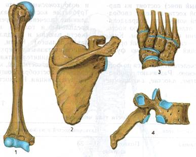
In humans, the anatomical and functional features of the limbs have developed under the influence of upright walking and labor. During the process of anthropogenesis, the forelimbs lost their importance for movement (locomotion) and turned into upper limbs. Their structure was mainly influenced by labor, under the influence of which the upper limb and especially the hand adapted to labor activity, turned into an organ of labor. The human hand is capable of not only grasping an object, as is the case in various animals, but also clasping it. Grasping is ensured by opposition thumb the remaining fingers of the hand. The lower limbs are adapted for movement and support of a vertically located body. This explains the structural features of the lower extremities: their massiveness, the appropriate location of the arch to the support. The foot has largely lost its grasping function. Despite the functional differences, the upper and lower limbs have a common structural plan. The skeleton of each limb is divided into the skeleton of the belt and the skeleton of the free limb. The upper limb includes the skeleton of the shoulder girdle and the skeleton of the free upper limb; the lower limb has a skeleton of the pelvic girdle and a skeleton of the free lower limb. Both belts are connected to the body.a) Skeleton of the upper limb:
on each side there are bones of the shoulder girdle (scapula and clavicle) and bones of the free upper limb (humerus, forearm and hand bones). Bones of the shoulder girdle: *Scapula - a flat triangular bone located on the back side of the chest in the superolateral part of the body at the level of ribs 2-7, connected to the spinal column and ribs with the help of muscles. The scapula has two surfaces (costal - anterior and dorsal - posterior), three edges and three angles. The shoulder blade connects to the collarbone. *The collarbone is a C-shaped, curved long bone that connects to the sternum and ribs.Bones of the free upper limb: * Humerus- refers to long bones, it has a middle part (diaphysis) and two ends (upper - proximal and lower - distal epiphyses). *The bones of the forearm are the ulna, radius, also long bones; accordingly, they are distinguished between the diaphysis, proximal and distal epiphyses. *The hand consists of small bones of the wrist, five long bones of the metacarpus and bones of the fingers. The bones of the wrist form an arch, concavely facing the palm. In a newborn they are just beginning; gradually developing, they become clearly visible only by the age of seven, and the process of their ossification ends much later (at 10-13 years). By this time, ossification of the phalanges of the fingers ends. 1 finger is of particular importance in connection with the labor function. It has great mobility and is opposed to all other fingers.
b) Skeleton of the lower limb:
each side includes the bones of the pelvic girdle (pelvic bones) and the bones of the free lower limb (femur, leg bones and foot). The sacrum is connected to the pelvic bonesBones of the pelvic girdle: * Pelvic bone consists of three bones - the ilium (located in the upper position), the ischium and the pubis (located at the bottom). They have bodies that fuse with each other at the age of 14-16 years in the acetabulum area. They have round depressions into which the heads of the femoral bones of the legs enter. Bones of the free lower limb: * Femur- the most massive and longest tubular among the long bones of the skeleton. *The bones of the lower leg include the tibia and fibula, which are long bones. The first one is more massive than the second one. *The bones of the foot are formed by the bones: tarsus (proximal part of the foot skeleton), metatarsus and phalanges of the toes. The human foot forms an arch that rests on the heel bone and the anterior ends of the metatarsal bones. There are longitudinal and transverse arches of the foot. The longitudinal, springy arch of the foot is unique to humans, and its formation is associated with upright walking. The weight of the body is evenly distributed along the arch of the foot, which is of great importance when carrying heavy loads. The arch acts like a spring, softening the shock of the body when walking. The arched arrangement of the foot bones is supported by a large number of strong articular ligaments. With prolonged standing and sitting, carrying heavy loads, or wearing tight shoes, the ligaments are stretched, which leads to flattening of the foot, and then they say that flat feet have developed. Rickets can also contribute to the development of flat feet.
Human skeleton(ancient Greek “dried”) - the totality of the bones of the body, the passive part of the musculoskeletal system. The name refers to the ancient method of making a skeleton - drying it in the sun or in hot sand.
The adult human skeleton contains about 206 bones, of which 33-34 are unpaired, the rest are paired. 23 bones form the skull, 26 - the spinal column, 25 - the ribs and sternum, 64 - the skeleton of the upper extremities, 62 - the skeleton of the lower extremities.
The bones of the skeleton are formed by bone and cartilaginous tissues, which are classified as cartilaginous tissues. Bones consist of cells and intercellular substance.
In adults, the skeletal-to-body mass ratio remains at 20% throughout most of their lives. In the elderly and old, this figure decreases somewhat. A dry, macerated (sequentially defatted, bleached, dried) human skeleton weighs 5-6 kg.
The hyoid bone, the only bone not directly connected to the others, is topographically located in the neck, but traditionally belongs to the bones of the facial part of the skull. It is suspended by muscles from the bones of the skull and connected by the pharynx.
There are also bones that do not belong to the skeleton. 6 special bones (three on each side) located in the middle ear; The auditory ossicles are connected only to each other and participate in the functioning of the hearing organ, transmitting vibrations of the eardrum to the inner ear.
Functions of the skeleton.
I. Mechanical:
support (formation of a rigid osteochondral skeleton of the body to which muscles, fascia and many internal organs are attached);
movement (due to the presence of movable joints between the bones, the bones work as levers driven by muscles);
protection of internal organs (formation of bone receptacles for the brain and sensory organs (skull), for the spinal cord (spinal canal));
spring (shock-absorbing) function (due to the presence of special anatomical formations that reduce and soften shocks during movements: the arched structure of the foot, cartilaginous layers between the bones, etc.).
II. Biological:
hematopoietic (hemopoietic) function (hematopoiesis occurs in the bone marrow - the formation of new blood cells);
participation in metabolism (it is the repository of most of the body’s calcium and phosphorus).
Skeletal structure.
The human skeleton is structured according to a principle common to all vertebrates. The bones of the skeleton are divided into two groups: the axial skeleton and the accessory skeleton. The axial skeleton includes the bones that lie in the middle and form the skeleton of the body; these are all the bones of the head and neck, spine, ribs and sternum. The accessory skeleton consists of the clavicles, scapulae, bones of the upper extremities, pelvic bones and bones of the lower extremities.
Axial skeleton
Scull- the bone base of the head, is the seat of the brain, as well as the organs of vision, hearing and smell. The skull has two sections: the brain and the facial.
Rib cage- has the shape of a truncated compressed cone, is the bone base of the chest and a receptacle for internal organs. Consists of 12 thoracic vertebrae, 12 pairs of ribs and sternum.
Vertebral column, or spine- is the main axis of the body, the support of the entire skeleton; The spinal cord runs inside the spinal canal. It is divided into cervical, thoracic, lumbar, sacral and coccygeal sections.
Accessory skeleton
Upper limb belt- provides attachment of the upper limbs to the axial skeleton. Consists of paired shoulder blades and clavicles.
Upper limbs- are maximally adapted to perform work activities. The limb consists of three sections: the shoulder, forearm and hand.
Lower limb belt- provides attachment of the lower extremities to the axial skeleton, and also serves as a container and support for the organs of the digestive, urinary and reproductive systems.
Lower limbs- adapted for support and movement of the body in space in all directions, except vertically upward (not counting jumping).


