How does a child behave during hypoxia? Treatment and diagnosis of fetal oxygen starvation. Drugs for oxygen deprivation
A woman preparing to give birth to a child can often be “scared” by various incomprehensible symptoms and diagnoses associated with pregnancy. After all, the main concern of every woman during this period is a proper pregnancy and, of course, the health of the unborn child.
That is why such a diagnosis as fetal hypoxia, or even sometimes just a suspicion of its presence, can cause real panic in the expectant mother. Of course, every woman wants to give birth to a healthy and strong baby.
Most events that cause suffocation occur in the womb; Because of this, it is important to have good prenatal care to identify problems that may be related to choking; for example, pregnancy-related hypertension problems, intrauterine growth retardation, placental problems, etc.
Cost-effective and affordable interventions can avoid maternal and perinatal mortality. Neonatal prognosis is closely related to maternal health care, and it has been estimated that 70% of fetal and newborn deaths can be prevented by maternal intervention. Possible long-term complications range from mild motor deficits to spastic quadriplegia with or without movement disorders, seizures, cognitive changes, neurosensory changes, and feeding problems.
But even if the doctor observing the course of your pregnancy suspects you have symptoms of hypoxia, you still shouldn’t despair. After all, all the emotional experiences of the mother are reflected in the child. So, what is fetal hypoxia and how does it manifest itself.
Fetal hypoxia, or so-called oxygen starvation of the fetus, develops if during pregnancy there is an insufficient supply of oxygen through the placenta and umbilical cord.
It is important to determine what degree of encephalopathy each strangulation patient has, since moderate scores have a normal prognosis; as opposed to those who have major changes or die or have severe serious long-term neurological complications. Other indicators of a poor neurological prognosis include: the presence of persistent or difficult to control seizures; abnormal neurological examination within a week of life; Difficulty feeding, which requires a nasogastric tube for a long time.
Of course, the first signs of fetal hypoxia during pregnancy can be detected by yourself. expectant mother. But this can only happen in the second half of pregnancy, when the baby begins to move. For more early stages During pregnancy, fetal hypoxia may not manifest itself in any way. The expectant mother, even with her presence, can feel great.
IN lately The role of imaging studies such as brain magnetic resonance in making long-term prognoses is being explored. Information Need Strategies need to be developed to improve neonatal and fetal prognosis. The first step to achieve this goal is "useful" information on perinatal mortality in all regions of Mexico, and this includes suffocation. It is necessary to determine which population has the best perinatal prognosis by comparing it with other populations and identifying the excess deaths that would be preventable if all pregnancies had the same opportunities as the population with the best reproductive prognosis in our country.
At later stages of pregnancy, a woman needs to pay attention to how actively the baby moves. As a rule, the child’s active movements can be divided into periods. That is, when the child is active and the woman feels him moving for at least a couple of minutes, and then the baby calms down for a while - this is one period. Then, after about an hour, the baby again begins to actively move in the mother’s stomach and calms down again - this is the next period. There should be at least ten such periods of movements per day.
With this strategy, areas requiring more intervention are identified to improve reproductive prognosis. You can't create strategies if you don't know the problems. There is currently a system for registering vital statistics. This is why it is necessary to have reliable, complete and timely statistics about the births that have occurred in the country, which allows us to have data about the mother, the newborn, the place of occurrence, the person who attends the birth and who confirms it. In this sense, the initiative to create a Certificate of Birth birth.
Many women are concerned about too much physical activity and sometimes the so-called “hiccups” of the baby, and they consider this a sign of hypoxia. But doctors are inclined to believe that reducing the number of movements of the child is much more dangerous. And it is precisely the decrease in the number of movements of the child that serves as the main sign of oxygen starvation of the fetus or hypoxia.
That being said, we have made great progress and it is expected that the entire Republic will use these certificates. Unfortunately, there is currently no “link” or connection between a birth certificate and a death certificate in Mexico. This does not allow specific mortality to be determined by birth weight, which is important for understanding the problem and creating intervention strategies. This is why it is necessary to establish a reliable and systematic perinatal surveillance system, with a system for analyzing information based on weight and age at death, allowing comparisons to be made to identify areas with excess mortality.
The child does not have enough oxygen and freezes for a long time. It is these symptoms that should alert the expectant mother.
The doctor observing you can also identify the first signs of fetal hypoxia during regular scheduled examinations. A symptom such as hypoxia can be clearly indicated by a decrease in the fetal heart rate. Or their uneven character, acceleration and deceleration of the heartbeat, disruptions in rhythm.
Birth weight is an important predictor of neonatal death. It is also clear that at lower birth weights and gestational age there is a greater need for neonatal resuscitation and a greater risk of asphyxia. Age at death is very important because it determines at what point in life death occurs. If, on the other hand, it occurs in the first 7 days of life, in addition to the analysis above, you should check temperature control, infections, nutrition, etc.
Systems approach for multifactor problem. Perinatal mortality is the result of a complex interaction of medical and social factors. Formal health care system: all health care facilities, private or public. Intersectoral system: includes systems that indirectly intervene in health, such as education, transport system, etc. Informal sector and community: these are families, the population, midwives. . The informal sector and community are an important target for reducing strangulation deaths.
In the second half of pregnancy, the baby's heartbeat can already be clearly heard through the abdominal wall. Of course, only a doctor can hear the baby’s heartbeat using a special obstetric stethoscope. When conducting routine examinations, based on regular observations, the doctor can identify symptoms of fetal hypoxia quite early and prescribe the appropriate treatment to the woman.
In Mexico, about 90% of births occur in institutions and the rest in homes. Pilot projects have shown that training midwives in neonatal resuscitation reduces deaths due to strangulation. Likewise, the meta-analysis also showed that this intervention improved perinatal survival and reduced mortality due to asphyxia. In addition, families should be trained to recognize obstetric emergencies and visit units promptly to seek care.
The formal healthcare system is basic. It is important to ensure adequate quality of medical care. Structures, processes and results must be reviewed. Currently, there are programs for neonatal resuscitation, treatment of high-risk newborns, neonatal stabilization, etc. Which should be distributed to personnel responsible for the delivery of infants. It is important that health authorities monitor such training. An obstetrician must be able to recognize and treat emergencies, and this includes performing emergency caesarean sections when indicated, the presence of blood, managing and recognizing hypertensive crises, etc. Close communication between gynecologists, pediatricians and neonatologists should be encouraged, and guidelines for perinatal management are created by consensus among them.
In addition, various deviations in the formation of the placenta can serve as an indirect sign indicating the presence of fetal hypoxia. A placenta that is too thin or, on the contrary, too thick and inappropriate for the duration of pregnancy, may lead the doctor to suspect the presence of fetal hypoxia. 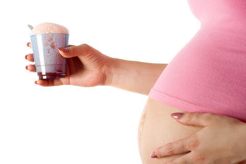
Also important is an intersectoral system that guarantees vehicles and roads for isolated communities will enable mothers to reach adequate care units. This system is especially needed in rural areas of Mexico. However, even in large cities, it is important to establish a transport system for high-risk mothers and newborns, allowing them to be transported to specialized centers; Without this measure, it is impossible to reduce newborn mortality.
Global efforts to prevent suffocation. A significant proportion of choking cases can be identified and treated in a timely manner. There are, however, some that are difficult to prevent or predict, such as acute umbilical cord problems. In these situations, the mother may notice decreased or absent fetal movement, which usually means that neurological damage to her baby has already occurred. These situations are currently unpredictable and difficult to prevent.
The beginning of placental abruption or its premature maturation is also a bad signal. Indeed, in these cases, the placenta is not able to provide the fetus with sufficient oxygen supply. Such symptoms of hypoxia can also only be identified by a doctor, based on the obtained ultrasound report of the pregnant woman.
With such signs of fetal hypoxia, a woman often requires observation of the course of pregnancy in a hospital setting.
This need has been identified by authorities around the world, which is how it was created by the Global Pediatric Research Program, which is conducting a "global crisis" analysis of this major public health problem. The team was formed by a group representing Salvando La Vida de Regien Nacidos; National Institute children's health and development; World Health Organization; International Pediatric Association; Canadian Newborn Network, members of various pediatric societies around the world and other associations or institutions.
If necessary, the doctor can give a referral for studies such as cardiotocography (CTG) or fetal Doppler. There is no need to be afraid of this. These procedures are absolutely painless and safe for the child. Such studies allow you to track the work of the child’s heart, as well as see whether the blood supply to the placenta and umbilical cord is normal.
Main conclusions. It should be remembered that not all birth sites have the ability to accept gasometry to detect the acidity of the fetus or newborn. Strategies must be created to prevent intrauterine asphyxia and fetal death. It is extremely important to promptly treat injurious asphyxia. train personnel intended to participate in childbirth. Records must be implemented to “fairly” measure the problem: its prevalence, risk factors, short-term and long-term effects, etc. There is a need to maintain close communication between all health care workers, join forces and create strategies to reduce or prevent perinatal asphyxia.
- It is necessary to have a universal definition of asphyxia.
- Long-term follow-up for neurological problems with choking is necessary.
Causes
So what can trigger hypoxia or oxygen starvation in the fetus? Every pregnant woman should know these reasons, because the sooner the presence of hypoxia is determined, the sooner the necessary measures can be taken to reduce it negative influence on the development of the child.
Most cases of fetal hypoxia during pregnancy are attributed to associated factors, and in to a lesser extent the development of hypoxia depends on hereditary predisposition.
Perinatal asphyxia is a major public health problem worldwide. In Mexico, it is responsible for the majority of newborn deaths. Unfortunately, over the past three decades, we have not seen a significant reduction in mortality due to this cause in our country or in other developing countries. To reduce infant mortality, it is necessary to develop strategies to prevent perinatal asphyxia by identifying and promptly treating conditions that affect the well-being of the fetus.
Researchers, clinicians, epidemiologists, gynecological obstetricians and neonatologists from around the world must join efforts to prevent and promptly treat perinatal asphyxia, as well as to accurately record this problem and its consequences.
Chronic diseases in a woman during pregnancy can also provoke the occurrence of fetal hypoxia: gestosis, diabetes mellitus, hypertension, bronchial asthma, anemia, various lung diseases and much more.
Also, fetal hypoxia during pregnancy can be caused by a woman staying in a stuffy room for a long time or maintaining an uncomfortable position for a long time. If a woman often lies on her back during pregnancy, the uterus can compress the inferior vena cava, which also causes symptoms of hypoxia. Therefore, pregnant women are not recommended to sleep lying on their back. Naturally, lying on your stomach is also not acceptable for pregnant women. Chronic fetal hypoxia during pregnancy can be observed if the expectant mother smokes during pregnancy.
Neuropathy and neonatal encephalopathy in Kathmandu, Nepal: assessing the contribution of asphyxia to perinatal mortality in a low-income urban population. No more screaming at birth: Global estimates of labor and delivery of the intrauterine newborn. Essential newborn care: report of a technical working group. Promoting newborn health around the world. Maternal and child health: are you ready? South Asia to change? Designing and using qualitative research to improve newborn care practice: A guide for program managers. The continuous value of the Apgar score for the evaluation of newborns. A look through Virginia's eyes. Committee on Fetus and Newborn, American Academy of Pediatrics and Committee on Obstetric Practice, American College of Obstetricians and Gynecologists. Apgar score use and abuse. American Academy of Pediatrics and American College of Obstetricians and Gynecologists. Recommendations for perinatal care. Effects of corticosteroids before preterm delivery: a review of evidence from controlled studies. Beneficial influence combined use of prenatal corticosteroids and postnatal surfactant in preterm neonates. "Newborns" of the world in the world, Hill, K. "Reducing perinatal and neonatal mortality." Special report on research in the field children's health, vol. 3, no. What supplies require the presence of pediatricians? World Health Organization, Child Health and Development: Health of the Newborn, Geneva, Switzerland: World Health Organization, Best evidence currently available: review of the literature on umbilical cord closure, immediate and delayed cord tightening in infants born between 24 and 32 weeks: a pilot randomized trial Controlled Study Placental Transfusion: Umbilical Cord Healing and Preterm Infants Delayed cord healing improves the hematologic status of Guatemalan infants at 2 months of age. Effect of cord clamping time on iron status in infants at 3 months. What can a meta-analysis reveal about traditional childbirth education and pregnancy outcomes? Thyroid responses to light are intact, spontaneous eye movements occur, and the wrist-eye maneuver is complete. Pupil size varies, although pupils tend to be dilated and reactive in less affected infants and reactive and miotic in more affected children. Most infants exhibit hypotonia at this stage. Seizures occur between 6 and 12 hours of birth in 50-60% of those affected, most of them manifesting in subtle movements, such as conjugate gaze deviation or sucking associated with altered consciousness. Premature neonates may exhibit decerebrate reactions that may mimic generalized tonic seizures. During this stage, the level of consciousness changes in a variable manner, with severely ill infants remaining dull or comatose, and less severely ill infants often demonstrating a degree of alertness that is more apparent than is actually the case and is not accompanied by fixation or visual monitoring. The attacks are intense and there are periods of apnea, agitation and weakness. Status epilepticus may occur, requiring aggressive and urgent treatment. Apnea and agitation may occur. The level of consciousness in severe cases deteriorates further and a deep stupor or coma occurs. Shields can be attached to the light in the middle position or extended. Those who die from hypoxic-ischemic encephalopathy do so more frequently during this period. Newborns who survive to this period usually improve within a few days to a few weeks; eating disorders are common and associated with problems with absorption, swallowing, and tongue movements. General hypotonia of the extremities is common, and some may exhibit hypertension, especially involving the basal ganglia. Neurological examination of a newborn differs from that performed in other age groups although it includes a detailed physical examination, it must also include analysis of primitive reflexes and requires knowledge physical changes observed in premature newborns and newborns They were exposed to asphyxia in different periods time, with their recovery and complications. The newborn examination should be done in a quiet place, with light and at an appropriate temperature to ensure that the newborn is comfortable after removal of clothing. Clothes should be removed slowly and gently, the diaper will only be removed while the area is being assessed. Ideally, the test should be carried out on the second or third day of life, a few hours after feeding and with the child not swimming or hungry. Important Steps in the study: observation. The study should begin with observation; it is a mistake to start manipulating a child without an appropriate period of observation. It is necessary to look for the presence of congenital anomalies such as defects midline skull, face, palate and spine. Abnormalities of the trunk, limbs and skin are easily detected. Changes in skin pigmentation are important because of the common ectodermal origin of the skin and nervous system. It is also important to observe limb position to assess passive muscle tone. Symmetrical movement of the limbs with slight flexion of the joints of the elbows and knees with spontaneous opening and closing of the arms is normal in newborn terms, while keeping the elbows stuck to the bed with little movement of the arms. hands may be normal until 32 weeks of pregnancy. In general, there is enough distance to go two fingers between the examination tray and the patient's neck, while larger distances may indicate hypertonicity of the cervical extensors and shorter distances may represent hypotony; the first - as chronic asphyxia, and the second - as data from acute asphyxia in the first hours. Asymmetrical mobility and asymmetrical handgrip should always be considered abnormal. Head. The head assessment includes external characteristics such as head circumference, shape and skin characteristics. Presence in the skin should be limited to the presence of masses, skin lesions such as congenital vascular malformations. It is normal to observe some bulging of the anterior fontanel with crying. The chain perimeter is a measure that informs us about the intracranial volume of both the brain and the cerebrospinal fluid; to a lesser extent, this measure depends on the subdural and subarachnoid space or intracranial blood volume. The reduced head circumference in the first days of life of a strangled child should make us suspect cerebral pathology before asphyxia. There are changes such as craniosynostosis, which is the premature closure of cranial sutures, which can affect one or more sutures, causing irregularities and asymmetries in the shape of the skull, always causing deformity and not microcephaly, in this case always the rules of cerebral and non-cranial pathology. The anterior fontanel is palpable at birth, sunken or flat; should be assessed with the child seated; bulging usually occurs during crying or when there is an abnormal increase in intracranial pressure; Its size ranges from 1 to 3 cm at its largest diameter, and its closure occurs between 10 and 20 months. The fontaneller works synchronously with the impulse. At birth, the posterior fontanel is punctate and open at birth, when intracranial pressure increases, it closes completely at 6 weeks of life. An important fact When analyzing a child with macrocephaly or microcephaly, it is to measure the cephalic perimeter of the parents, since common familial variations exist. During the first days of life, a tight fontanel, a period when the baby is choking, is usually a sign of a poor prognosis and usually represents severe cerebral edema. Warning status. Changes in consciousness are identified by analyzing periods of anxiety and sleep. When a newborn reacts to external stimuli, such as a human voice, touch, light. Babies who sleep are easily awakened by external stimuli, and those who cry are easily soothed. Warning is one of the most sensitive functions that depends on the integrity different levels central nervous system. In the newborn, the state of alertness varies and depends on several factors such as gestational age and time since the last meal. At 28 weeks' gestation, alert periods are difficult to identify, with constant stimulation, eyepiece opening, and warning periods achieved within seconds; At 32 weeks of gestation, they experience spontaneous ocular opening and may experience periods of ocular rotation and periods of sleep. Examination of cranial nerves. At 26 weeks of pregnancy, newborns often flicker in response to light; at 32 weeks, close your eyes while you present a light stimulus; and at 37 weeks, the engineer looks at the light stimulus. To evaluate this cranial nerve, the pupils must exhibit the same response to stimulation in order to be symmetrical; bright light makes newborns blink or keep their eyes closed. When assessing the fundus of the pallid optic disc, lower vascularization is also observed than in older children. The student is greyish-white. 20% of retinal hemorrhages not associated with complications during childbirth or disorders of the central nervous system or neurological complications and completely eliminated are observed in 7 to 14 days. Detailed analysis and searching for specific retinal disorders associated with asphyxia, should be specifically examined by an ophthalmologist with experience in the field of child care and asphyxia of premature infants. Lack of eye movement in the direction expected results in altered midbrain level nuclei or pathways of these nerves. In term, a large object hitting the color can also be used in the form of a red circle or browser face, the target focus moving slowly into the newborn's field of view. Pupils are difficult to assess because the eyes are closed and resist forced opening. The corneal reflex should be studied, which is when the eyes are closed when the cornea is lightly tipped with a sock. Remember that the mouth is redirected to the opposite side of the facial palsy and that when it comes to closing the peripheral palsy the eye and the affected side where the patient remains open the eye is also affected. From 28 weeks of pregnancy the auditory nerve function is evident to provide a response to sound stimuli. To the extent that maturation increases, the response is most obvious, resulting in performance motor activity, changes in breathing patterns, opening of the mouth and eyes. To test hearing function, it must be done when the child is calm, without tears, without hunger, and to ensure that the ear canals are clear. One of the most common complications in patients is asphyxia of premature hearing loss, so it is necessary to check the integrity of the auditory pathway by performing auditory evoked potentials and otoacoustic emissions. Nutrition involves proper coordination of breathing, salivation and suction. Suction and salivation are properly coordinated from the 28th week of pregnancy, but breathing is not, so food is inconsistent at this age. Delay in the acquisition of these skills in newborns with asphyxia usually determines a poor functional prognosis for food and then for the tongue, a form of pseudobulbar palsy, which are the upper control centers that cause coordination failure. Neonatal depression central nervous system has a little weakness and a tear. During vigorous crying, the symmetry of the newborn's tongue and palate can be assessed. When studying these parameters, gestational age should be taken into account and assessment should be carried out in children early age over 24 hours of life. The first thing to assess is the newborn's resting position and spontaneous limb mobility. Although the tone is assessed, the newborn should be lying on his back with his head inward, so that the tonic neck reflex does not increase the tone unilaterally. A vertical suspension should be performed to determine whether the tone of the flexor limb is adequate and symmetrical, since in the presence of hypotonia, newborns in this position tend to slide between the arms of the browser. In the ventral perspective, the newborn's pendulum acquires a vertical position between the trunk and the head, while in the presence of hypertonia of the neck extensors, the head tends to exceed the trunk. Tone assessment is also performed from the heel approach to the ear, the hand on the contralateral ear or by measuring the flexion angles of certain joints like the wrist, ankle or popliteal angle. The position and tone depends on the gestational age of the maturity of the flow towards the rostral, at 28 weeks of gestation there is minimal resistance to passive movement of all limbs, at 32 weeks the flexor tone predominates in the lower extremities and at 36 weeks the flexor tone is also evident in the upper extremities; passive mobilization of the limbs in full-term infants is an obvious tone of the limb flexors above and below; Head position also depends on gestational age; in full-term infants, it shifts steadily to the right and is often less obvious during the first 24 hours of life. In newborn asphyxia, hypotension is often initially identified, which causes spontaneous mobility; the limbs typically hang by the Landau maneuver and remain attached to the bed when the supine position is reduced. Strength assessment should be taken into account in the movements of the limbs. During the first 4 weeks of life, sinuous movements predominate, from 4 to 12 weeks are characterized by restlessness and nervousness, from 8 to 12 weeks, broad movements and strength predominate. It helps to grasp the arms or legs and pull gently to measure the force exerted to bring the newborn's arm to a normal flexed position. Muscle stretch reflexes assessed in newborns are the thoracic, biceps, brachioradialis, patella and Achilles. Reflexes cause a reflex hammer in accordance with the size of the child, increase, and sometimes may be absent in healthy newborns, although they are always symmetrical and tears often increase them. The extensor response of the plantar should be symmetrical, although sometimes it is indifferent to the flexors during crying or sleep. Persistent plantar extensors after the child reaches an abnormal rise and a violation of the corticospinal route, as a completely asymmetrical response, and especially in combination with a violation muscle strength and tone. At the 28th week of pregnancy, only the opening of the arm is present; at 32 weeks, abduction and expansion upper limbs is present and at the 37th week flexion of the limb is already obvious. The asymmetrical arm is caught in a corticospinal injury. The reflection from 35 weeks of gestation becomes increasingly obvious during the month of life and disappears between 6 and 7 months. In addition to information about clinical-electroencephalographic correlations to induce or inhibit movement of stimuli, it is important to determine information about pathophysiology. The main types of neonatal seizures are shown in Table Table.
- Best practices: identifying and treating newborn asphyxia.
- Monitoring perinatal mortality.
- Pathophysiological approach.
- Early clinical manifestations From birth to 12 hours.
- The severely affected infant is in deep stupor or coma.
- There is usually intermittent breathing or breathing problems.
Hypoxia can also be caused by a number of complications during pregnancy. Such as:
- polyhydramnios or large amounts of amniotic fluid;
- pelvic diligence of the fetus;
- multiple pregnancy;
- disruptions in the uteroplacental blood supply;
- pathologies of placental development;
- fetal infections during pregnancy.
Consequences of fetal hypoxia
Unfortunately, fetal hypoxia during pregnancy can be both acute and chronic. And the severity of the manifestations of the consequences of hypoxia often depends on how rapidly the process develops.
For example, if we're talking about about premature ripening of the placenta or maternal smoking during pregnancy, the child constantly experiences a slight lack of oxygen. This is of course very bad, but does not pose an acute threat to the child’s life.
And the development of an acute process, as a rule, can lead to the most tragic consequences, including the death of the baby. This situation can occur if complete premature placental abruption occurs during pregnancy. The child stops receiving enough oxygen and may soon die. The development of this condition in a pregnant woman requires immediate medical attention. In case of acute hypoxia, a caesarean section is performed, only then is it possible to save the child.
Children who were exposed to hypoxia during pregnancy are often born with underweight or stunted growth, even if the pregnancy was full-term. Sometimes there is a developmental delay, increased nervousness of the child, and pathologies in the development of the cardiovascular and nervous systems.
Treatment and prevention
An acute attack of fetal hypoxia is sometimes impossible to predict and prevent. But chronic hypoxia, if detected early, can be treated and controlled. Of course, only if you strictly follow the recommendations of your gynecologist. The main advice to reduce the risk of fetal hypoxia is to avoid bad habits, if any. Also, the expectant mother should try to go for walks more. fresh air, move actively if your health allows, avoid long stays in stuffy rooms.
A necessary condition for a normal pregnancy is a proper diet. A woman needs to eat a varied diet so that the body receives a sufficient amount of vitamins and microelements from food. Also, it is necessary to include iron-rich foods in the diet, since iron deficiency anemia is one of the reasons for the development of chronic hypoxia during pregnancy. At the same time, try not to overeat, because overweight during pregnancy, it provokes the development of shortness of breath and difficulty breathing.
Try to attend scheduled obstetrician appointments in a timely manner and take all necessary tests so that the doctor can see the presence of any abnormalities early on. Consult your doctor if you have any unpleasant symptoms or suspect the development of hypoxia. And also tell your doctor if you have any chronic diseases that can cause fetal hypoxia.
To prevent the development of fetal hypoxia, pregnant women are often recommended to perform special breathing exercises. A good preventive measure is water aerobics. Of course, if you have no contraindications for such activities. 
Sometimes, to saturate the blood of a pregnant woman with oxygen and to prevent the development of fetal hypoxia, the use of so-called oxygen cocktails– special water or phytosolutions enriched with oxygen. Another way to treat hypoxia is to prescribe sessions in a pressure chamber for a pregnant woman. Staying in a pressure chamber is prescribed in a certain course and helps to saturate the mother’s blood with oxygen, and therefore a better supply of oxygen to the fetus.
And most importantly, even if you are directly faced with such an unpleasant diagnosis as fetal hypoxia, first of all try to remain calm and not get nervous. After all, any of your experiences are acutely transmitted to your baby.
Intrauterine hypoxia, oxygen starvation, lack or lack of oxygen in the fetus during pregnancy are different definitions of the same pathology. The essence of which is quite clear from its name. Sadly, almost every 10th pregnancy occurs with intrauterine hypoxia.
The baby in the womb does not breathe on its own. Along with nutrients, oxygen is supplied to him through the placenta via the umbilical cord. If there is insufficient oxygen supply through the uterus-placenta-fetus system, the child experiences all the “delights” of its deficiency. It is customary to distinguish:
- spicy;
- chronic
The first most often develops during childbirth. The second is detected during the gestational period, and worries the baby while he is in the womb.
During fetal hypoxia, it is customary to distinguish between threatening oxygen starvation and confirmed hypoxia. First, this is a condition in which there are no obvious symptoms of pathology yet, but hypoxia can be assumed if fetal maturation is impaired. The latter can be determined using ultrasound and CTG (from 30 weeks). It is divided into three stages according to severity:
- mild degree leading to;
- medium, which causes metabolic disorders in the fetal body;
- severe causes irreversible changes at the cellular level.
The pathology becomes chronic when diagnosed untimely. At any stage of detection, chronic oxygen starvation of the fetus requires treatment.
Acute hypoxia can develop at any stage of pregnancy; more often this condition is typical for the final stage of gestation. Usually it occurs due to tight repeated entanglement of the umbilical cord around the body or limbs of the fetus and bending of the umbilical cord. As a result, blood stops flowing to the fetus.
The same effect can occur when the umbilical cord vessels thrombose or nodes form on it. Treat acute hypoxia not possible. It is urgent to remove the fetus to save its life. 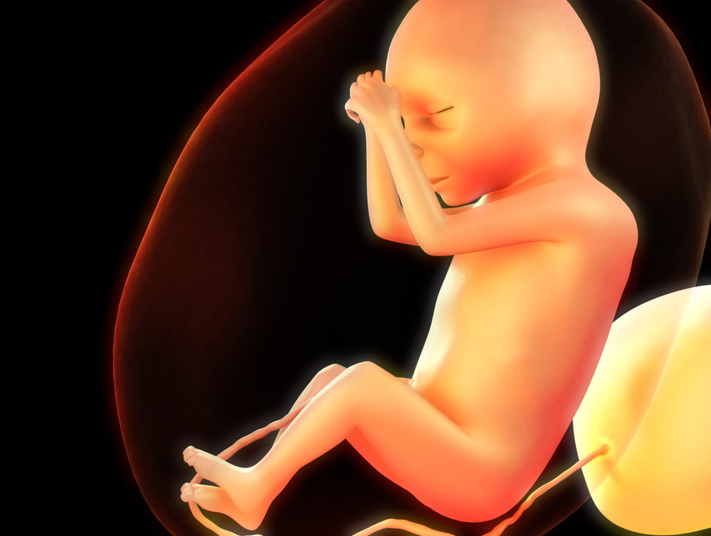
Causes of lack of oxygen
Acute hypoxia often develops as a result of delivery pathology: weak labor activity, prolonged compression in the birth canal. Repeated tight entanglement or too short an umbilical cord, which interferes with the expulsion of the fetus, contributes to its getting stuck in the birth canal and the development of hypoxia.
A blood clot or umbilical cord knot leads to the same result. The use of anesthesia sometimes leads to oxygen starvation of the baby.
There can be a large number of reasons for the development of chronic hypoxia. All factors that provoke chronic lack of oxygen can be divided into three large groups:
- related to the woman’s health;
- associated with the state of the uterus-placenta-fetus system;
- associated with external harmful factors and bad habits.
The first group includes: anemia, kidney disease, pathology of the respiratory system, heart and vascular diseases, endocrine disorders present in the mother. The most common causes are: anemia and diseases of the bronchi or lungs.
Anemia or anemia is a pathology caused by a lack of hemoglobin, the main “transporter” of oxygen. With anemia, hypoxia develops in both the mother and the fetus.
Respiratory diseases of an inflammatory and allergic nature (rhinitis, hay fever, chronic bronchitis and asthma) lead to impaired nasal breathing or a decrease in the supply of oxygen from outside for other reasons. 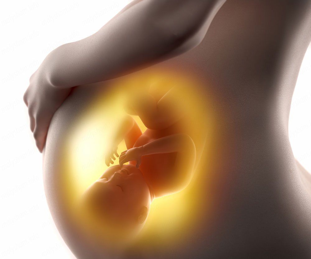
The second group includes pathologies affecting the uterus, placenta, umbilical cord and the fetus itself. Hypertonicity of the myometrium, fetoplacental insufficiency, chorion or placental abruption, abnormalities in the structure of the umbilical cord, intrauterine infection of the fetus, polyhydramnios and oligohydramnios cause inadequate oxygen supply to the fetus.
Postmaturity and compression of the umbilical cord vessels as a result of repeated entanglement can cause hypoxia during pregnancy.
Environmental factors can also cause pregnancy pathology and hypoxia. Such unfavorable factors include:
- passive and active tobacco smoking;
- environmentally unfavorable region of residence or harmful production conditions;
- prolonged or frequent stay in an area exposed to smoke;
Separately, we can highlight psychological factors, which are not the last link in the chain of development of oxygen starvation in the fetus. Severe negative stress leads to spasm of blood vessels, including placental ones, and a decrease in their bandwidth, and hence a decrease in oxygen transport.
Signs of intrauterine hypoxia
In the early stages of pregnancy, it is impossible to independently determine that the baby is suffering from oxygen deficiency. During this period, the woman does not yet feel the movement of the fetus and oxygen deficiency can only be detected during examination. In the second half of gestation, when the expectant mother clearly feels the beating of the fetus, she should be alerted to:
- increasing its activity (increasing the frequency of movements and the force of blows);
- reducing the number of movements to 10 times a day or less and weakening the tremors.
This characteristic features lack of oxygen in the fetus. Many expectant mothers are at a loss as to what exactly should be considered a sign of oxygen deficiency: hyperactivity or apathy of the baby growing in her womb. In fact, there is no contradiction. 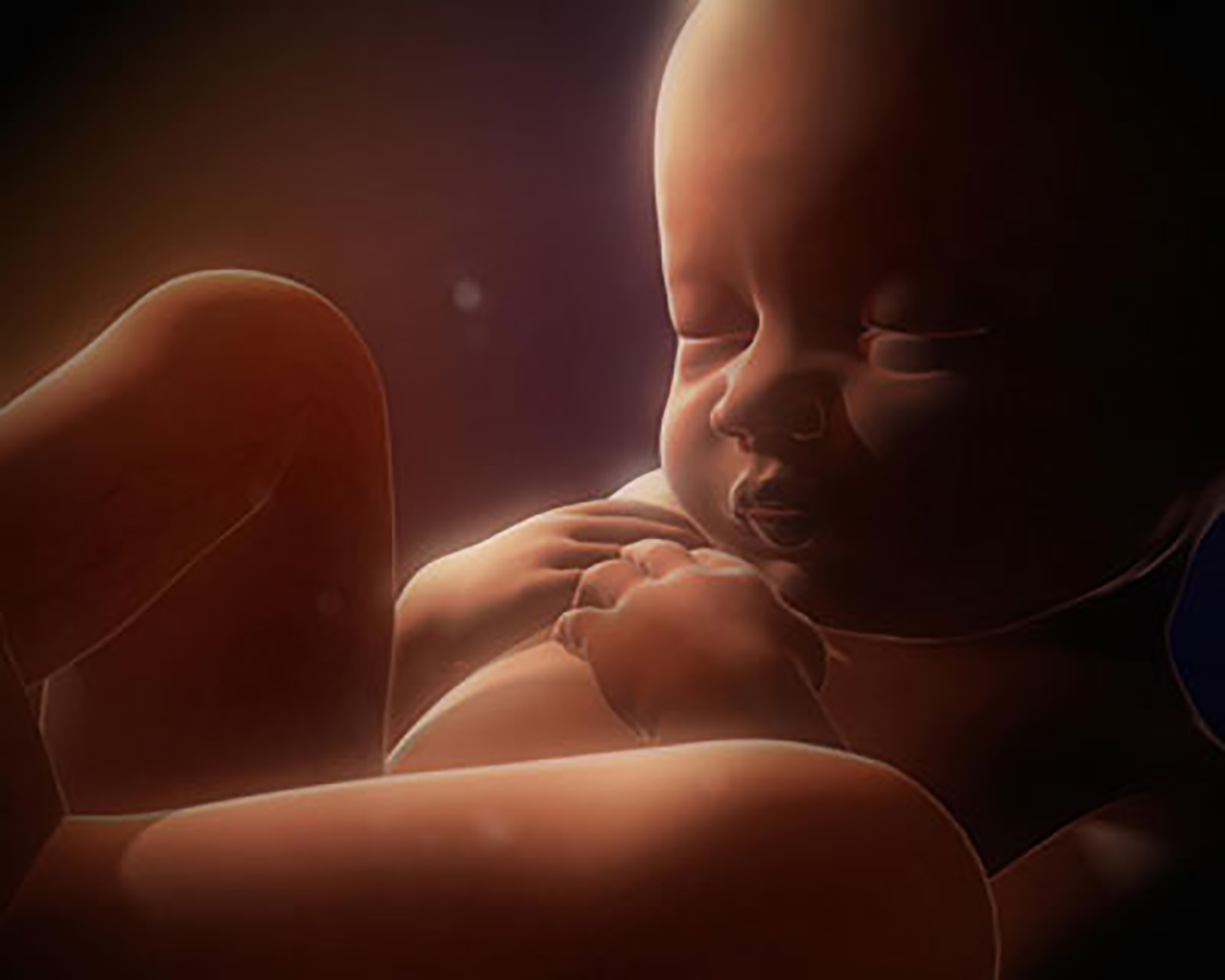
At the first stage of oxygen starvation, the fetus, like any human being, is characterized by motor excitation. But the longer this deficiency continues, and the more significant it is, the slower the fetal movements become, and weakness engulfs it. Both the number of movements and their intensity decreases. In this case, you should immediately consult a doctor.
It is worth noting that the daily activity of the fetus must be taken into account, since it must sleep and rest. Temporary cessation of movements after a period of activity is not considered a pathology.
Threat of oxygen starvation
Chronic oxygen deficiency most often causes fetal malnutrition and neurological disorders in the newborn. In addition, after birth the child may experience thermoregulatory disorders and weak nonspecific resistance, anemia.
With a constant lack of oxygen, brain cells suffer, so over time, at the stage of socialization, a child may develop impairments in cognitive functions (memory, attention, thinking).
Acute hypoxia that develops during childbirth can become a factor provoking pneumonia. The risk of developing cerebral palsy and damage to nerve tissue increases. In some cases, intestinal ischemia and necrotization of its tissues develop. 
Treatment and prevention
Chronic oxygen starvation, depending on the severity, can be treated at home or in a hospital setting. Therapeutic tactics depend on the cause of the disease. In case of iron deficiency anemia, the mother may be prescribed iron supplements. Women with uterine hypertonicity may be prescribed antispasmodics.
When diagnosing oxygen starvation of the fetus, all expectant mothers are recommended to rest, vitamin supplements, and oxygen cocktails.
In order to prevent hypoxia, a woman is recommended to carefully prepare for conception, conduct healthy image life during the period of bearing a baby, take active walks (if health allows), get proper rest, and avoid stressful situations. To prevent nutritional anemia, it is worth introducing vegetables and fruits into your diet; pomegranate and its juice, as well as liver, are especially effective.


