How much does an abdominal CT scan with substance cost? Abdominal CT scan: what is it? Computed tomography of the abdominal cavity: what it shows
CT abdominal cavity– a diagnostic method that describes the condition internal organs abdominal cavity (together with vessels and abdominal lymph nodes) and retroperitoneal space layer by layer with a step size of 0.5 to 10 mm.
Computed tomography provides an informative, highly accurate three-dimensional image, revealing small changes in organs that are invisible with other studies. High scanning speed makes it possible to obtain results within 2 hours, and low radiation exposure to the human body expands the indications for use. With CT it is possible to differentiate tissues due to the difference in their density with an accuracy of 0.5%.
Contraindications to CT scan of the abdominal cavity
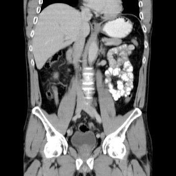
Paralytic ileus Paralytic ileus can have several causes, including the use of certain medications, as a reaction such as abdominal inflammation or after abdominal surgery. No sudden changes in calibration are observed. Mechanical ileus. Mechanical ileus can also have various causes. In mechanical obstruction, the intestinal loops proximal to the mechanical obstruction dilate and collapse distally. It is important to detect this sudden change in calibration and examine it in several directions to find out exactly what is happening here.
A computed tomography scan of the abdominal organs includes the following:
- gastrointestinal organs (pancreas, liver, large and small intestines, stomach, spleen, gall bladder);
- retroperitoneal space (kidneys, adrenal glands, blood vessels, urinary system, lymphoid tissue).
CT scan describes the structure of parenchymal organs, its painful changes, and the patency of tubular organs.
The adhesion itself is almost never visible. Sometimes several cycles are involved. This occurs in a "closed loop" obstruction. In this situation, the intestines are removed in two places, for example. by adhesion, internal hernia or torsion. Intestinal loops in a closed circuit expand and connect as if at the site of obstruction. Adhesion causes venous collection in the mesenterium, characterized by increased mesenteric density. Torsion, also called wolf torsion, can occur in both the small intestine and large intestine.
Indications and contraindications
In the colon, the sigmoid is usually involved. 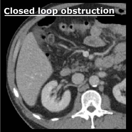
Closed loop small bowel obstruction secondary to adhesion. Intestinal perforation is characterized by extraluminal, “free” gas in the abdominal cavity. Free gas is usually located around the perforation site, allowing some conclusions to be drawn regarding the location of the perforation. There is also free fluid in the peritoneal cavity due to leakage of intestinal contents or peritoneal irritation.
CT scan of the abdominal cavity can show volumetric processes of pathological changes (size of the inflammation, its boundaries and extent of spread) and failure in the functionality of organs, namely:
- foreign bodies;
- damage to the lymph nodes;
- hemorrhages and injuries;
- vascular changes and atherosclerosis;
- cysts, malignant and benign tumors;
- abscesses;
- blood diseases;
- hepatitis, cirrhosis, liver hepatosis;
- kidney and gallstones;
- echinococcosis;
- congenital anomalies.
Indications for a computed tomography scan of the abdominal cavity may also include:
Gastric perforation with free gas. Hint: Free gas can be easily detected in most cases by examining the abdomen in a “lung setting.” The same patient as in Fig. 37, in mild conditions. With intestinal ischemia, the blood supply to the intestine is limited. This may be the result of an occlusion in the arterial supply, for example. blood clot, but it can also develop as a complication of mechanical intestinal obstruction. In addition, thrombosis in the superior or inferior mesenteric vein, in the absence of adequate collateral information, can cause increased retrograde pressure, thickening of the intestinal wall, and ultimately decreased intestinal perfusion.
- preparation for surgery;
- resolving the issue of operability;
- acute conditions and pain of unknown etiology;
- chronic digestive and urinary disorders that cannot be explained by other diagnostics;
- obstructive jaundice and sudden weight changes;
- acute abdominal injuries;
- suspicion of a mass formation in the abdominal cavity;
- treatment control.
This is the most reliable way to identify cancer at an early stage. The pictures show the size, boundaries of the inflammation zone, local metastases and the degree of tumor invasion into neighboring organs and lymph.
Intestinal ischemia is usually characterized by decreased strengthening of the intestinal wall and “pneumatosis intestinalis,” defined as gas in the intestinal wall. In severe cases, this gas can enter the mesenteric or even portal veins through the intestinal wall. In the absence of these characteristics, intestinal ischemia cannot be excluded.
Such products include
Intestinal ischemia of the ascending colon secondary to thrombosis in the superior mesenteric vein. Severe intestinal ischemia with pneumatic bowel and portal gas. Prokop; Spiral and multilayer computed tomography of the body. . Scanning is performed without preparing the patient in emergency situations, such as trauma, differentiation of ischemic and hemorrhages, as well as diagnosis of subarachnoid hemorrhages. In addition to the above conditions, the patient should always be on an empty stomach if intravenous contrast media is to be administered.
Using an abdominal CT scan, you can accurately diagnose complex diseases such as:
- mediastinal tumors - lymphoma and lymphogranulomatosis;
- hemangioma and hepatocellular carcinoma;
- hemochromatosis;
- pheochromocyotoma;
- kidney cancer, hydronephrosis;
- polycystic disease;
- nephroptosis;
- hemoblastosis;
- abdominal aortic aneurysms, stenoses, kinks of blood vessels;
- portal and mesenteric thrombosis;
- cholangitis, diverticulitis, appendicitis;
- intervertebral hernia or pinched nerve;
- gallbladder pathology;
- pancreatitis;
- adenoma or cyst of the adrenal gland.
With contrast
Native CT is performed without the use of contrast. But most often more accurate data is needed, then a targeted CT scan of a specific abdominal organ using contrast is prescribed.
The scanning protocol must include fragments smaller than 1 mm, which are only available in multi-layer scanners. Older types of CT scanners, in which these options are not available, do not allow staff to obtain optimal images of all structures of the inner and middle ear. This allows assessment of the auditory ossicles, cochleae, hemispherical canals, internal and external acoustic ducts, mastoid pneumatization, congenital anomalies, post-traumatic lesions, and both benign and malignant growth processes.
Contrast is the introduction of an x-ray contrast agent inside (the amount is proportional to the patient’s weight) to increase the accuracy and reliability of diagnosis due to the fact that the contrast agent enters the painful tissues through the blood and makes them brighter and clearer in the image.
To get more detailed information For parenchymal (solid tissue) organs such as the liver, pancreas and kidneys, oral contrast is used. To study blood vessels, contrast is administered intravenously. There are situations when it is most advisable to administer contrast through an enema. Bolus intravenous contrast is administered at a predetermined rate using a special automatic injector.
Features of preparation for CT with contrast when examining various organs
With the exception of post-traumatic conditions and the search for foreign substances in the eye sockets, scanning is performed using a contrast agent. The scan is performed without a contrast agent, unlike diagnosing patients with a tumor disease. During the scan, the patient usually lies on his stomach with his head tilted back.
Patients with suspected primary or secondary sinus cancers are scanned using a contrast agent and in such cases should be present for the scan on an empty stomach. This method has been applied, for example, in the assessment of cerebral blood flow. It allows visualization of the penumbra and ischemic areas before the formation of irreversible morphological lesions in it, namely myocardial infarction. Along with the increase in the number of slices in CT scanning, there is an increase in the volume of brain volume in which blood flow is examined.
During the scan, the patient will have to lie quietly on his back for several minutes, breathing evenly, possibly holding his breath for a few seconds. The uniform is loose, metal accessories will need to be removed. Native diagnosis will take up to 15 minutes, with the introduction of contrast – about 30 minutes. The likelihood of adverse reactions such as nausea, anxiety, fever is observed in only 1% of cases out of 100.
This allows for a better assessment of ischemia and its range. These examinations should be performed after intravenous administration of contrast material, which should be performed before the patient has eaten. Neck imaging is performed to evaluate lymph nodes, inflammatory and neoplastic infiltrates, and connections to arterial and venous vessels, respiratory tract or salivary glands.
Abdominal CT scan: what does the scanning procedure show?
Technological progress has made it possible to study vessels of ever smaller diameter. Currently, multi-slice tomographic scanners make it possible to scan such large anatomical areas as arterial vessels of the lower extremities, hip joints and the terminal part of the abdominal aorta. To depict blood vessels, the blood circulating in it must be mixed with a contrast medium; Because large amounts of this medium must be managed, the study uses non-ionic media, which reduces the risk of complications.
A radiologist is best able to interpret finished images, especially in controversial issues. Specialists in a narrow field - surgeons, oncologists, gastroenterologists can also decipher. It usually takes no more than 1.5 hours to evaluate the results and prepare a conclusion.
This allows you to evaluate parenchymal organs - liver, spleen, pancreas and kidneys. In addition, the gallbladder and extrahepatic and intrahepatic bile ducts are also studied. This method allows you to check the range of inflammatory and neoplastic infiltrates in the walls of the stomach, small and large intestines, adrenal glands and assess the likelihood of relapse at the site of tumor removal. Proper patient preparation is always required. Intestinal loops should be filled with a 1-2% aqueous solution of iodine, an ionic contrast medium that allows differentiation of the inner wall of the intestine, its wall and structures adjacent to the intestine.
Preparation
Preparing for an abdominal CT scan requires careful planning and compliance. At the time of tomography, the gastrointestinal tract should be as free from food as possible.
Diagnostics is carried out on an empty stomach, so if it is prescribed:
- in the morning - a light liquid dinner is allowed;
- for lunch - 5 hours before you can afford a very light breakfast;
- in the evening - the patient takes a light liquid breakfast and refuses lunch.
Since food passes through the esophagus for about 2 days, during this period it is necessary to avoid foods that cause bloating and complex foods: alcohol, carbonated drinks, fermented milk products, fermented foods, apples and cabbage, legumes, yeast products, hard and indigestible foods. food.
Before the scan, the patient remains on an empty stomach for 5-6 hours. The amount of orally administered fluid and the time spent administering contrast depend on the indication for the examination. With helical multislice computed tomography scanners, dynamic multiphase scanning has become standard. Perfusion studies in the liver and pancreas are something of a novelty.
It is based on checking these organs in both the arterial and venous phases, as well as examining the liver and the portal. The opportunity for dynamic research arose in connection with the introduction of spiral machines, especially multilayer ones. Diagnosis of tumors: undifferentiated liver disease, primary hepatic neoplasms, metastases Post-procedural: complications, perfusion disorders Vascular: portal vein thrombosis, vascular anatomy, Osler's disease Other: abscess, trauma Quantitative measurements: hemosiderosis, liver volume and tumors. Therefore, it was impossible to assess wall thickness, any change in bowel position or change in the width of their lumen.
Immediately before the procedure, a cleansing enema is performed, or in the evening before the test day, you can cleanse the intestines with Fortrans solution (oral administration of a laxative solution). The bladder may be slightly full.
From the evening until the start of the CT scan, you need to drink up to 4 liters of still water in equal portions, with 2 ampoules of 20 ml of pharmaceutical Urografin 76% or Triombrast 60% diluted in it. If a change in the source of inflammation is suspected, the doctor may prescribe intravenous administration of Omniopak (50 ml).
Virtual colonoscopy allows you to evaluate the inner surface of the intestinal walls. Another prerequisite for obtaining a clear image of the inside of the intestine is the complete emptying of food debris from the intestinal lumen. The patient must be prepared as for a traditional colonoscopy. However, there is a possibility that in the near future, contrast media will be available that will bind to the chemist and allow it to stand out in the picture. The quality of this method increases with the reduction in examination time due to peristalsis and the need to hold the breath.
Regardless of the area of examination, plenty of still water at room temperature is required after a CT scan.
Contraindications
CT is a safe examination; the radiation dose of the longest diagnostic procedure does not exceed the average dose of a conventional X-ray of the gastrointestinal tract.
The main restrictions for its implementation are: large body weight (over 120 kg), pregnancy, restless state of the subject.
The exam can only be carried out using multi-slice tomography. Other groups of recommendations include assessing the range of infiltration in a neoplasm of the esophagus, lesions of the mediastinal lymph nodes, pulmonary embolism, lesions after radiation therapy, neoplastic infiltrates arising from the pericardium or heart, and trauma chest. To obtain intense enhancement of the heart and large vessels in the mediastinum, intravenous administration of a contrast agent is required, which makes it possible to differentiate pathological changes from the normal state of the organs.
Relative contraindications in each individual case may be:
- age up to 14 years, only if it is impossible to establish a final diagnosis using other studies;
- acute kidney inflammation;
- Breastfeeding is not recommended within 24 hours after a CT scan with contrast;
- CT with contrast is not performed in cases of severe diabetes mellitus;
- allergic reaction to contrast agents(iodine and derivatives);
- in case of severe diseases of the liver and cardiovascular system, do not use contrast;
- blood diseases;
- taking adrenergic blockers and other incompatible drugs;
- if in less than 7 days the patient underwent an X-ray of the gastrointestinal tract with barium contrast.
Preliminary examination
Before undergoing a CT scan, you may need to undergo other examinations, such as an abdominal ultrasound, colonoscopy, FGS of the stomach and duodenum, conventional x-rays, or a referral from an oncologist to draw up a report.
Tumor diagnosis: lung and mediastinal tumors, secondary tumors, tumor assessment, assessment of tumor progression. Detection and Quantification: Occult inflammatory process, decay, asbestosis, silicosis, edema, bronchiectasis. Previous site: biopsy, bronchoscopy, bronchoalveolar lavage, differentiation of lung lesions from pleural lesions. High-quality computed tomography - Diffuse lung diseases.
Detection of contained lesions in the lung parenchyma Morphological characteristics Quantification of lesions in the lung parenchyma Location of the lung or bronchoalveolar lavage before open biopsy. Another sign is pathology of the intervertebral disc and the surface of adjacent vertebral bodies. Patients do not need to fast. The scan usually does not require contrast material.
Before performing a computed tomography scan with contrast, it is necessary to consult an anesthesiologist, clarify contraindications with a doctor, and obtain written consent for the administration of a contrast agent.
When performing a CT scan of the kidneys, adrenal glands, bladder and ureters, the patient must have fresh (2-3 days) results of a biochemical blood test for ALT, AST, urea and creatine on hand
The first group of indications consists of examinations without contrast media and is aimed at assessing the bone structure in the case of Paget's disease, fibrous dysplasia, post-traumatic lesions or degenerative joint disease, in case of an ambiguous clinical and radiological picture in a plain x-ray image. Another group of indications, in addition to the evaluation of bone structures, includes pathology of the soft tissues around the bones and inflammatory and neoplastic infiltrates, both those that extend from the bones to the soft tissues and vice versa.
The doctor's referral for a CT scan will indicate the research method, area of examination, presumptive diagnosis and purpose of the diagnosis.
Examination price
The price of diagnostics using a computed tomograph depends on the complexity of the study and the location of the inflammation. The average cost of an abdominal CT scan is determined by the availability and cost of its components and consumables:
- post-diagnostic consultation;
- consultation and management of an anesthesiologist during CT;
- recording images on electronic media;
- tomography;
- administration of contrast;
- administration of contrast intravenously;
- bolus intravenous contrast;
- cost of contrast agent;
- printed photographs, research package and duplicates;
- intravenous anesthesia;
- puncture (puncture) of the organ being examined for diagnostic and treatment purposes.
Primary native CT scan of the abdominal cavity usually costs from 3,500 rubles in Moscow. The average cost of a CT scan with contrast includes only basic services and ranges from 6,000 rubles. When studying cancer patients, the required accuracy increases to a cutting step of 0.5-3 mm and proportionally affects the price.
The abdominal cavity is one of the most important areas human body, uniting organs that are responsible for the processes of digestion, hematopoiesis, the formation of immunity and perform other life support functions. To diagnose diseases in this area, a computer scan is performed - a modern, highly informative diagnostic method. What is the preparation for an abdominal CT scan, and what should the patient know?
Abdominal Anatomy
The abdominal cavity is the space that is located under the diaphragm and is separated from it by the azygos muscle. Conventionally, this space is divided into three sections (“floors”) and contains the following vital organs:
Often a CT scan is performed not only of the abdominal cavity, but also of the retroperitoneal space. What is the retroperitoneal space? This is the area adjacent to the peritoneum, which contains:
- The pancreas is an organ that takes part in the digestion of carbohydrate and fatty foods and regulates carbohydrate metabolism.
- The kidneys are responsible for the function of urine formation.
- Adrenal glands – endocrine glands regulating hormonal levels.
- The ureters, which connect the kidneys and bladder.
- Part of the intestine.
- Lymph nodes.
Thus, this is a vital area of the human body, and its examination and timely treatment of detected pathologies can significantly improve the quality of life, and sometimes prevent the development of fatal diseases.
What can a computed tomography scan of the abdominal cavity show?
CT scanning of the retroperitoneal space and peritoneal organs visualizes the organs listed above, as well as vessels and abdominal lymph nodes. The procedure is carried out to obtain images in three-dimensional format and in high resolution. From a diagnostic point of view, it is very important that CT of internal organs allows you to obtain a layer-by-layer image of the organ due to the fact that images are taken from different angles.
What does a CT scan show? A CT scan is recommended to detect pathological processes, including inflammation and the appearance of tumors. This method allows you to see:
- Presence of foreign bodies.
- The appearance of abscesses.
- Formation of neoplasms.
- Hemorrhages.
- Vascular disorders.
- Changes in the functioning of the lymph nodes.
In addition, CT scans of the abdominal cavity and retroperitoneal space are used in preparation for operations and when deciding on their feasibility. This is an indicated method for disorders of urinary function and digestion, if other diagnostic methods have not brought results. CT scan of the retroperitoneum and peritoneum is performed after injuries and after operations when it is necessary to check their effectiveness.
CT scan of the retroperitoneum and peritoneum is performed after injuries and after operations when it is necessary to check their effectiveness.
CT scan of the abdomen and retroperitoneum can be performed with. It uses an iodine-containing drug. This is the procedure most often used:
- To obtain more accurate information about the condition of parenchymal organs, oral administration is used.
- If RCT of the abdominal cavity is performed, including to study the condition of blood vessels, intravenous contrast is used.
CT scan of internal organs, unlike MRI, is x-ray method research, and during it the patient receives a certain dose of radiation. Let us list what contraindications CT of the retroperitoneal space and abdominal cavity has:
- Pregnancy. Breastfeeding is considered a relative contraindication: after the examination you need to take a break for at least a day.
- Age up to 14 years. In this case, CT is performed only on the condition that making an accurate diagnosis using other methods is impossible.
- Presence of barium mixture in the stomach. This situation arises if the patient has already undergone a CT scan of the retroperitoneal space and peritoneum no later than a week before.
- If the study involves the use of contrast, contraindications are allergies to iodine, dysfunction thyroid gland, heavy diabetes mellitus, renal failure, and serious illnesses liver and heart.
How to prepare for a CT scan?
Preparation for a CT scan of the abdominal cavity and retroperitoneum and peritoneum involves examining the condition of the organs involved in the digestive process. The organs that are located in this area are located very closely, and therefore the slightest change in size or displacement of at least one of them can lead to the fact that the result will not correspond to reality.
In this regard, preparation is required before an abdominal CT scan. What can you do before going for a CT scan?
Important! You cannot eat at least 6 hours before the test!
In this regard, it is worth preparing and planning your day and diet depending on what time the CT scan is scheduled:
- If the diagnosis is carried out in the morning, you can have a light dinner.
- If the procedure is scheduled for the day, a light breakfast is allowed, but no later than 6 hours before the start.
- If the study is carried out in the evening, breakfast is allowed, but it is better to refuse lunch.
It is worth preparing for the study by conducting certain types of tomography; you may need to take some tests. For example, blood biochemistry for creatinine and urea - if contrast is supposed to be administered to a patient with renal pathologies.
When choosing where to conduct research, you should pay attention to the qualifications of specialists working in one or another medical center. The accuracy of the results will depend, firstly, on the correct preparation and behavior of the patient during the scan. Not lower value The doctor has the ability to evaluate the results obtained. This can also be done by the doctor who issued the referral for a CT scan, but radiologists themselves have the most experience in interpreting images.
-
 April 17, 2015Chicken broth Calorie content of pork broth
 -
 April 17, 2015Chicken, tandoori masala, oven
 -
 April 17, 2015Recipes with chicken and tandoori sauce


