Computed tomography radiation safety. X-ray examinations and CT scans: necessity and danger
Computed tomography (CT) is a research method that is based on obtaining a series of x-rays (slices) of the body and their computer processing to obtain a detailed image internal organs.
Computed tomography should not be confused with magnetic resonance imaging (MRI), which uses a different imaging principle.
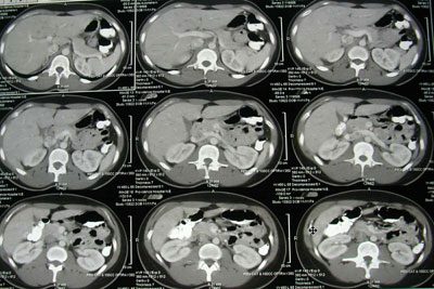
With a CT scan, the doctor receives many sections of the patient’s body, on which one can see certain anomalies. The sections can be placed at a distance of only one millimeter from each other, so the image is very detailed, and the doctor will not miss even the smallest defect or tumor. In addition, CT images can be combined by a computer, resulting in a three-dimensional image of any part of the body (organ, vessel, bone).
CT is much more convenient and informative than traditional x-rays.
Why is a CT scan done?
CT has a wide range of uses in medicine, including in examining patients who have been in a serious accident and have suffered internal trauma. A CT scan will immediately help determine where the patient has fractures or other problems.Diagnosis of fractures or tumors of the musculoskeletal system.
. Detection of internal injuries and bleeding after injury.
. Determining the location of a blood clot, source of infection or tumor.
. Diagnosis of many diseases of blood vessels, heart, lungs, liver, etc.
. Monitoring during procedures (biopsies, radiation therapy etc.)
Risks associated computed tomography:
1. Exposure to radiation.
During a CT scan, the patient is exposed to radiation, and in a higher dose than during a conventional x-ray examination. This exposure is associated with a very small chance of developing cancer in the future.
However, CT has clear benefits that outweigh the potential risks. Doctors use minimal power to keep the radiation exposure as low as possible. Newer CT scanners emit less radiation than older models. If you are concerned about radiation, talk to your doctor about alternatives to CT scanning.
2. Harm during pregnancy.
You must inform your doctor about your pregnancy before having a CT scan. In this regard, the doctor may recommend another examination method, such as magnetic resonance imaging or ultrasound. The latter method is absolutely safe for the fetus.
3. Reaction to contrast agent.
In some cases, the doctor has to use a special contrast agent, which is administered intravenously or given to the patient to drink before performing a CT scan. Therefore, rare allergic reactions to the contrast agent, including anaphylactic shock, are possible. But in most cases, allergic reactions to contrast are mild, limited to skin rash, redness and itching. Be sure to tell your doctor if you have had an allergic reaction to contrast media in the past.
Preparing for a CT scan
Preparing for a CT scan depends on which part of the body will be scanned.The patient may be asked:
Take off some clothing and put on a hospital gown.
. Remove all metal objects and jewelry from the body.
. Stop eating some time before the scan.
Contrast agent for tomography
Contrast agents are needed to “highlight” certain structures on x-rays. The contrast agent blocks X-rays and lights up white in pictures, which helps highlight blood vessels, intestines or other parts of the body. This will enable the doctor to recognize certain defects that would go unnoticed without contrast.Methods of administration contrast agent some:
Orally. If the doctor plans to examine the esophagus or stomach, then the contrast must be taken in the form of a special solution. It may have an unpleasant taste.
. Parenterally. Contrast agents are often injected into the bloodstream through a vein in the arm. This is done before studying the gallbladder, urinary tract and blood vessels. During the injection, the patient may experience a feeling of heat at the injection site, as well as a metallic taste in the mouth.
. Rectally. To visualize the intestines, a contrast agent is administered rectally. This procedure is associated with discomfort. There must be enough contrast agent so that it covers the intestinal walls and makes them clearly visible on the pictures.
Preparing young children for a CT scan is a very difficult task. Before the scan, your doctor may recommend a sedative to help your child remain quiet and not twitch during the procedure. Any, even minor movements can affect the results of a CT scan because the clarity of the images is lost.
What should I expect during the procedure?
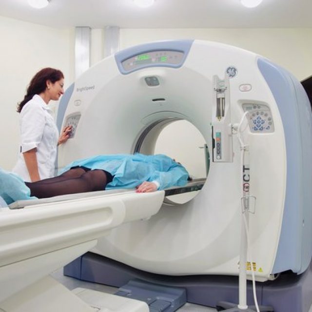
Computed tomography is a completely painless procedure. And with the use of modern machines, CT scans have become very fast, often requiring only a few minutes for the entire process.The CT scanner looks like a huge donut. The patient lies down on a table that fits into the “hole” of this donut. Special belts and pillows help the patient maintain the desired position and not move during the procedure. During a CT scan of the head, a special “cap” may be placed on the patient’s head, which will help keep the head absolutely still.
The table can move during a CT scan, and the machine will rotate around the patient, taking pictures of the body under different angles. Each rotation can provide several images with the thinnest sections of the body. The patient will hear clicking and other strange sounds - this is normal.
The doctor conducting the study will be in the next room behind the monitor. He will be able to communicate with you through an intercom, so if any problems arise, you can report them. Your doctor may ask you to hold your breath to take pictures of certain areas.
After the tomography, you can immediately return to your normal rhythm of life. If you receive contrast dye, your doctor will give you special instructions. In some cases, he may ask you to stay in the hospital to make sure you feel well after the procedure. After the scan, it is recommended to drink plenty of fluids so that the kidneys remove the contrast from the body faster.
Images obtained from a CT scan are usually stored on storage media as a computer file. A radiologist interprets these images and sends them to your doctor.
MSCT is an abbreviation for the name of a relatively new medical method of examining the body - “multi-layer (or multi-slice) computed tomography.”
This diagnostic technique is based on unique abilities X-rays. To carry it out, special equipment is used, which is both a source of X-ray radiation and a means of perception and analysis of rays passing through body tissue.
Due to the fact that in the process of passing through tissues with different densities, radiation wastes its power, fixing it at the output makes it possible to create an image of internal organs and environments. The resulting image is used by doctors for diagnostic purposes.
How does MSCT differ from CT?
The main difference between MSCT - multilayer computed tomography and CT - conventional computed tomography - lies in the special capabilities of the equipment used.
Devices used for MSCT latest generation, in which one stream of X-rays is captured by several rows of detectors. This allows you to simultaneously obtain up to several hundred sections and significantly reduces the duration of the study: in one rotation of the emitting element, an entire organ is scanned. The clarity of sections is increased and the number of defects associated with the movement of internal organs is minimized.
The high speed of MSCT makes it possible to study not only the structure of organs, but also the processes occurring in them, causing minimal harm to the patient: the dose of radiation he receives is reduced by three times compared to conventional CT.
Which is better, MSCT or MRI?
The fundamental difference between MSCT and MRI is that the first technique is based on the properties of X-ray radiation and involves exposing the patient to X-rays. In the second case, diagnostics are carried out using an electromagnetic field, which has a more gentle effect on the human body.
However, MRI has a much wider list of contraindications - it cannot be used if the patient has metal prostheses, implants and tattoos applied with metal-containing dyes. Fear of closed spaces and mental disorders. Additionally, MRI is a more expensive procedure and most clinics only use it for certain indications.
How is MSCT examination performed?
To perform a conventional MSCT, the patient is placed on a special couch equipped with a lift, which is easily moved into the capsule of the device emitting X-rays. Maximum time stay in the device is several tens of minutes, but the radiation time does not exceed a minute.
The procedure is not accompanied by unpleasant sensations and does not require special training or compliance with the instructions of medical personnel.
To improve image quality, an iodine-containing contrast agent is injected into the patient’s body before MSCT. Before examining the organs of the digestive system, it is offered to drink, and when examining tissues and blood vessels, it is administered through a vein. In this case, the study is carried out several tens of seconds after the administration of contrast and generally differs from standard multislice tomography only by an increase in duration.
How often can MSCT be done?
The frequency of MSCT does not have this of great importance, as the amount of radiation received during the diagnostic process. The recommended threshold for exposure to radiation received during preventive examinations by the Chief Sanitary Doctor of Russia is 1 mSv (millisievert) per year, while a dose of 5 mSv is considered the most harmless.
The average radiation dose received during multislice tomography ranges from several fractions of hundredths to several tens of millisieverts. Each dose received is recorded in a special radiation exposure sheet. The possibility and necessity of each subsequent examination is determined individually, based on general condition patient and the need to obtain new diagnostic data.
How to prepare for MSCT?
A day or two before multispiral tomography of internal organs, foods that cause severe gas formation should be excluded from the diet.
A few hours before the upcoming study, food intake is stopped. Liquid ( clean water or water with a contrast agent dissolved in it) is taken evenly, in small portions.
Before examining the pelvic organs, it is necessary to empty the intestines, if necessary, by performing an enema.
The upcoming MSCT of the head or osteoarticular apparatus does not require special preparation.
How long does an MSCT study take?
The unique capabilities of the equipment used for MSCT can significantly reduce the duration of the study.
Thus, conventional multislice computed tomography lasts from several minutes to several tens of minutes, depending on the area and depth of the area being examined.
The duration of the examination procedure using a contrast agent can be increased to an hour. In some cases, the administration of the contrast agent begins several hours before the examination, then the entire diagnostic process takes several hours.
What is the radiation dose for MSCT?
The radiation dose that a patient receives during MSCT (multispiral computed tomography) is determined by the area and depth of the tissues to be examined, the type of device used and the examination technique.
As a rule, the radiation exposure when examining one anatomical area is within the limits of 3-5 mSv (millisieverts). A lower load is associated with the examination of bones and joints (a dose of about 0.0125 mSv), and a higher load is associated with the diagnosis of internal organs. During an in-depth examination of organs chest or abdominal cavity these values can increase noticeably, reaching several tens of millisieverts.
How much does MSCT cost?
The price for multislice computed tomography is determined not only by the pricing policy of the medical institution, but also by the quality of the equipment used during the study, the level of complexity of the procedure, as well as the qualifications of the medical personnel.
In 2015, the average cost of studying one anatomical area using MSCT falls within several (2-3) thousand rubles. The cost of studying blood vessels, especially with the use of a contrast agent, is estimated much higher - it is about 10 thousand rubles. A heart examination is estimated even higher, the cost of which reaches 17-18 thousand.
Patients often ask questions about whether CT scanning is harmful to health. People clearly have grounds for concern, since computed tomographs use X-ray radiation that is unsafe for human health to obtain images. This does not mean that it is necessary to abandon CT scanning altogether. This means that the choice of examination method must be taken into account the age of the patient and the radiation dose that the patient may receive during the procedure.
Is CT scanning harmful for adults?
For patients over 18 years of age, maximum permissible radiation doses per year have been established that will not cause damage to human health. Exceeding these doses is extremely undesirable and is allowed only in cases where the examination must be performed for health reasons. For example, in case of traumatic brain injury, when it is necessary to find out whether there is an intracranial hematoma.
Radiation doses are taken into account not only from CT scans, but also from fluorography, x-rays of bones and joints, and other procedures that use x-rays.
CT is not a diagnostic method that can be used in the first place, and especially for preventive purposes. If it is possible to obtain the necessary information using ultrasound or other accessible and safe examination methods, then it is better to give preference to them.
Why is CT scanning harmful for children?
The sensitivity of a child's body to the damaging effects of X-ray radiation is 5 times higher than in adults. The more actively a child grows, the more actively the cells of his body divide and the higher the risk of developing cancer in response to X-ray radiation.
To reduce the risk of developing cancer, it is necessary to ensure that the personnel operating the CT scanner adjust the machine settings to take into account the weight of the small patient. After all, what smaller child, the easier the X-rays pass through his body. Accordingly, the lower the dose load for conducting an informative examination.
How to minimize the harm of CT?
There are several simple rules, compliance with which will help reduce the dose of x-ray radiation that the patient receives during the examination:
- conduct an examination of the problem area of the body using other diagnostic methods, such as ultrasound, FGDS and others, and only if simple and safe methods do not provide the necessary information, take a referral for a CT scan;
- do not undergo examination to make sure that you do not have a disease: “preventive” CT examinations can do more harm than good;
- It is necessary to examine only the problem area of the body; “just in case” you should not expose nearby areas to X-rays;
- It is best to carry out the examination on a multispiral tomograph, since it gives half the radiation dose compared to a spiral one;
- The more modern the tomograph model, the better.
Harm from the use of contrast agents
We should not forget about the possible adverse effects of contrast agents on human health. Used for CT scanning contrast agents based on iodine can cause allergic reactions and a whole series side effects.
Computed tomography (CT) is the most modern method X-ray examination. Such an examination allows you to take a whole series of x-rays of the patient’s body, after computer processing of which you can obtain a detailed flat or three-dimensional image of the internal organs. Is CT scanning harmful to health? This is the question patients ask when there is a need for prompt and detailed diagnosis of internal organs after an accident or sharp deterioration health status.
This fear of patients is quite understandable. Firstly, due to the disease they have weak immunity and poor health. Secondly, patients know that there are a large variety of research and diagnostic methods, such as ultrasound (US), magnetic resonance imaging (MRI), fluorography, radiography and fluoroscopy (with layer-by-layer scanning and obtaining a series of slice-by-slice images). distance of a few millimeters - computed tomography).
Modern research methods help make accurate diagnoses and clarify previously made diagnoses in a short time, but they also have their own specific disadvantages. Some equipment based on these methods is completely safe, but very expensive and removed from potential patients, some have a number of contraindications for use, and some are harmful and even dangerous, if used incorrectly, for the patient’s health.
How does computed tomography work?
 Computed tomography refers to x-ray methods for examining patients, such as radiography, fluorography and mammography, and is based on the properties of ionizing radiation.
Computed tomography is performed on devices of various modifications, which have the appearance of a streamlined and convex ring, reminiscent of a flying space object. The tomograph also includes a retractable table, which smoothly feeds the patient under study into the apparatus to pass the patient through the entire system, including an ionizing emitter and a receiver of such radiation.
When a person passes through a computed tomograph, ionizing radiation is transmitted through the patient’s body, some of the radiation is absorbed by tissues, and such rays are then captured by several sensors. That is, the so-called transillumination of the patient’s internal organs occurs.
After receiving the stream ionizing rays the sensor system transmits information about the state of internal organs to the computer, with images displayed on the display of the diagnostic device. Scanning of the patient's body is performed layer by layer, every few millimeters, depending on the selected parameters. This allows you to get using computer programs both a detailed flat image of organs, areas of the musculoskeletal system, and a three-dimensional image after computer processing of slice images.
With the development of the medical industry, more and more advanced equipment is being developed that makes it possible to examine a patient in a matter of minutes with minimal consequences for health.
Received detailed graphic image the entire patient’s body can be enlarged or reduced in scale, studied in digital or paper form. This method of examination is prescribed by attending physicians to confirm and sometimes clarify previously made diagnoses, to conduct intermediate controls of the course of treatment and to diagnose possible pathologies in the early stages of development.
Features of the CT examination
 Computed tomography for restoring health is considered one of the progressive methods of examining patients. This method is an innovative x-ray method for diagnosing human diseases.
This method of examination differs from a conventional x-ray in that during scanning of the patient, not one picture is taken, but a whole sequential chain of pictures in the form of layer-by-layer sections of the patient’s body. The resulting images, after computer processing, can be studied both on the device’s monitor and on paper. The pictures are flat and volumetric images internal organs.
If it is not possible to examine the body using such harmless methods as ultrasound or MRI, then the attending physicians resort to CT scanning.
But we must remember that the operating principle of computed tomography is based on ionizing radiation, which in large doses can have a harmful effect on the human body.
An appointment for any examination of internal organs must be obtained from your attending physician. Before the examination begins, the tomograph staff provides a short briefing on the procedure and also performs a number of activities:
- The staff warns about the presence of a panic button designed to stop the examination of the body.
- To make this procedure easier, when the patient passes through the scanning chamber, specialists may not place pillows under the patient's head.
- Flow fresh air is directed at the patient using an auxiliary ventilator, which helps to distract the person being examined.
- During scanning, it is advisable to place the patient on his stomach.
- If possible, the attending physician refers the patient for examination using an open-type apparatus.
When examining small patients or patients with severe claustrophobia, healthcare workers suggest taking medicines sedative effect.
The impact of computed tomography on the patient’s health
 During examination using computed tomography, the patient receives radiation from 3 to 10 mSv. The radiation dose to a person depends on the size of the study area, the selected mode of operation of the device, that is, on the number of images in a layer-by-layer section and, of course, on the age and modification of the tomograph.
Scientists have calculated that, on average, one procedure on a CT scanner corresponds to the background radiation received by a person for 2 years everyday life. And radiation tends to accumulate in the body. Experts have also determined the maximum annual human radiation dose to be 150 mSv. However, cancer can appear and develop even at a dose of 50 mSv. Therefore, doctors recommend not to exceed the radiation dose of 50 mSv.
The harm of CT scans to the human body is obvious, but with the development of modern medical technology it can be minimized. It is important to adhere to certain rules.
How is CT more harmful than MRI?
 In order to answer the question of what is more harmful than MRI or computed tomography, it is necessary to consider the principle of the design and operation of the devices of such examination methods.
When examining a patient with a computed tomograph, his body is exposed to small doses of ionizing radiation. When this procedure is repeated, the radiation accumulates, so it has some limitations and contraindications. For example, examination in this way is prohibited for pregnant women.
In diagnostic devices based on MRI, there are no sources of radiation, and the patient during the examination is exposed only to a strong, but completely safe electromagnetic field. Therefore, with such devices it is possible to undergo repeated examinations without restrictions.
Clear, clear picture the condition of the patient’s internal organs or tissues is the dream of every doctor. It is this that helps to discard the subjectivity of the patient’s feelings and gives the opportunity to make a diagnosis not on assumptions, but on seeing the location of the real pathological process, its vastness and possible complications. That is why the development of diagnostic imaging methods has been and is priority direction medicine.
We recommend reading:X-ray: harm or benefit?
One of the first major breakthroughs in this area was the practice of X-ray examinations, and today it still occupies one of the key places in medical diagnostics. Despite the advanced age of the method and the emergence of new technologies, it helps save the lives of patients. However, X-rays pose a health hazard. Paradox? Remember the famous saying of Paracelsus: “Everything is poison, and nothing is without poison; Just one dose makes the poison invisible”? This also applies to X-rays in the best possible way.
Of course, modern X-ray technology has become much safer and more accurate, but it is still based on the same radiation, about the dangers of which more than one medical treatise has been written and many legends and myths have developed. Let's try to separate truth from lies and understand the question - why x-rays are harmful, how much you should be wary of x-ray diagnostic procedures and computed tomography.
X-ray research method
In relation to new methods, x-ray remains the most inexpensive, informative, takes little time and does not require a large staff of medical personnel. These circumstances determine its prevalence at the present time.
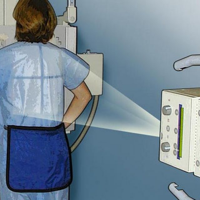 The method itself consists of passing x-rays through the body tissues and then recording:
- on a fixing film (radiography);
- on a black-and-white or color monitor (fluoroscopy);
There are many methods of X-ray examination. Each of them requires a certain time, amount of radiation, and multiplicity. It is necessary to understand that with each irradiation the patient receives a certain dose, which not only causes certain processes in the tissues of organs and systems, but can also accumulate. The doctor’s task is to minimize the existing harm from radiation exposure.
If dynamic monitoring of research data is required (for example, the passage of a barium mixture through the esophagus, intestines), then fluoroscopy is used. Despite the long-term exposure of the patient to radiation, the dose is much lower than with radiography, which takes an almost instantaneous image, but is significantly more powerful.
Sometimes fluoroscopic and radiographic methods are used simultaneously, which reduces the radiation dose to the patient, but at the same time provides more information
How can x-rays be harmful to a patient?
The danger of x-rays to human health is sometimes significantly exaggerated, but denying it is fundamentally wrong.
Harm is ionizing radiation that affects the human body with a certain intensity and time interval.
Radiation passing through body tissue:
- ionizes tissue molecules;
- causes a temporary change in blood composition;
- changes the structure of proteins;
- causes premature aging of cells;
- disrupts the normal process of maturation and life of body cells;
- promotes the development of cataracts;
- causes the degeneration of body tissues into pathological ones.
The result of these changes can be various diseases, among which malignant formations are possible. Therefore, it is very important when carrying out radiology diagnostics take all necessary precautions.
Important: Medical equipment uses low-energy X-rays, applied in short periods of time, and is therefore considered virtually harmless. The risk of a malignant tumor process increases on average by no more than 0.001% after a routine study. Electromagnetic waves emitted by an X-ray machine do not accumulate in the body.
Please note: It should be separately noted that different tissues of the body have different sensitivity to radiation.
Danger of X-rays for children
 The developing child's body, due to its imperfection, is more susceptible to negative influence ionizing radiation than an adult. The danger of X-rays for children lies in the possibility of malignant degeneration of cells entering the irradiation zone.
How younger age child, the higher the danger and the more often complications may occur.
In addition, in children the genetic component of protein molecules suffers, which can lead to the provocation of existing hereditary diseases.
Important: The answer to the question – is x-ray harmful to a child is clear: harmful. Therefore, each diagnostic procedure using radiation must have clear indications.
X-ray diagnostic methods and pregnancy
X-rays during pregnancy should be subject to even more stringent criteria, since exposure to rays on a developing child in utero is especially dangerous for him. If the doctor decides that this type of examination cannot be avoided, then he is obliged to take all precautions to minimize the harm of the procedure.
 This can be achieved:
- using the most radiation-friendly method;
- The study should be carried out with a reduction in the temporary load on the patient;
- use all necessary protective materials that can contain the level of radiation from the device;
- use modern X-ray equipment and materials.
Most often during pregnancy, an x-ray of a tooth/several teeth and a photograph of the jaw apparatus are used. This is due to the fact that the dental tissues of the expectant mother are exposed to caries in a more aggressive form.
But even in this case, x-rays should be done only when:
- resolving the issue of tooth extraction;
- purulent periodontitis;
- to check the quality of filling the root canal with filling material.
Please note: when ordering an X-ray examination, the patient is obliged to notify the doctor about the presence of an existing pregnancy. The doctor’s task is, if possible, to avoid using this diagnostic method or reduce its harm.
Comparative doses and risk assessment of radiological diagnostic methods
Speaking about radiation doses, it should be noted that three categories of data are subject to recording:
- absorbed dose - energy received per unit of human body mass;
- equivalent dose - absorbed dose multiplied by a coefficient, characterizing different degrees of damaging radiation;
- effective equivalent dose - calculated from the product of the equivalent dose with the sensitivity coefficient of different tissues.
Important: To assess the harm of ionizing radiation, we will be interested only in the EED - the equivalent effective dose, measured in mSv per unit of time (millisievert).
There are tables with a large amount of data, including comparative figures characterizing the average dose of radiation received. Based on them, you can calculate the expected amount of radiation during the upcoming X-ray examination.
We present one of them, the data is presented in μSv (microsievert, or 10 -3 mSv):

But it’s worth mentioning right away that all these data are approximate and, according to current radiation safety standards, limits for doses received by patients during the examination are not established. This is due to the fact that the minimum dose that can cause the onset of an oncological process in a person is considered to be 50 mSv/year, which is almost impossible to obtain using x-ray methods.
X-rays can pose a significant health hazard only to working personnel. The permissible dose in this case is up to 1000 mSv for the entire period of work.
You can get more information about X-ray radiation, the length of X-ray waves and the dangers of X-rays by watching this video review:
Features of computed tomography
Computed tomography differs from a conventional x-ray examination in that it takes not just a picture of a body area, but “layer-by-layer sections.”
This allows:
What are the harms of computed tomography?
Just like with a regular X-ray examination, the tissues of the human body remain exposed to radiation. This means that the risk of developing oncological processes remains.
-
 April 17, 2015"Marble" cupcake: recipes and cooking methods
 -
 April 17, 2015Recipe without sterilization with onion sautéing
 -
 April 17, 2015Lazy achma in a slow cooker
 -
 April 17, 2015Achma from lavash in a slow cooker


