Cervical region. Skeleton of reptiles.
The cervical section of the axial skeleton is a flexible single-arm lever. During its movement, it relies on that area of the neighboring thoracic, which coincides with the girdle of the thoracic limbs. The anterior end, which gives the greatest range of movement of the lever as a whole, articulates with the skeleton of the head, which is the reason for the diversity of the design of the cervical lever in terms of its length and mobility, as well as the strength of the muscles working on it.
The head, among numerous devices, also contains the initial section of the digestive organs, with which animals capture nutritional material; therefore, the entrance to oral cavity Herbivorous animals in a standing position must reach the ground. From this it is clear that the length of the neck in ungulates (with rare exceptions) must correspond to the length of the thoracic limbs, i.e. long limbs require also long neck(camel, giraffe), short limbs - short neck (pig).
The large length of the cervical lever in representatives of the class of mammals is not associated with an increase in the number of cervical segments, of which there are always seven, but depends entirely on the length of the supporting pieces, i.e., the bodies of the cervical vertebrae. In addition, the length of the neck in most terrestrial vertebrates is inversely proportional to the size (heaviness) of the head and neck muscles: the heavier the head and muscles, the shorter the neck and the less mobile in terms of freedom and ease of rotation in various planes, but it has enormous strength, and vice versa .
The shortening of the neck, noted as a secondary phenomenon in many aquatic mammals (whales, dolphins, etc.), is entirely explained by the need they have to have a strong connection between the head and the body to cut through the water when swimming (fish-like design).
The length of the cervical vertebral bodies of most terrestrial mammals, starting from the last (7th), gradually increases anteriorly up to and including the 2nd, and then sharply decreases at the 1st vertebra with the body absent.
The mobility of the articulations between adjacent vertebrae is generally small, but it increases significantly with the simultaneous movement of several segments, and the connection of the head of one vertebra with the fossa of the neighboring one (via intervertebral cartilage) allows movements in the cervical region to be made with a greater scope than in the thoracic and lumbar regions. The heads of the vertebral bodies of the neck are strongly convex and protrude in relief. In terms of swing force, the cervical lever is second only to the caudal section of the axial skeleton.
The neck, together with the head attached to it, undoubtedly plays a large role in the movements of the animal, contributing by its raising (or lowering) to the rapid movement of the center of gravity of the body, which is necessary to maintain its balance when pushing with the limbs (for example, in horses).
The mobility of the head near the 1st vertebra and the 1st vertebra near the 2nd, i.e., in the two front joints of the neck, has its own characteristics.
In the joint between the occipital bone and the 1st vertebra, mobility is quite developed, especially in the sense of flexion and extension within the midsagittal plane. The joint between the 1st and 2nd vertebrae allows significant rotation of the cervical lever around the longitudinal axis. Thus, already in these two joints there is a special modification of the mobility of the cervical spine, due to a modification of the connecting surfaces of the 2nd vertebra with the 1st and this latter with the occipital bone; Because of this, the very shape of these vertebrae is greatly changed. The originality of this form in comparison with the form of other cervical bells made it necessary to single out the 1st and 2nd vertebrae and give them their own names.
The first cervical vertebra is called the atlas. It is deprived of a body, which is replaced by a ventral arch (the rudiment of the body grows to the body of the 2nd vertebra at an early stage of development). The atlas has: a) a relatively large vertebral foramen, b) two wide lamellar transverse processes-wings of the atlas, c) clearly defined cravial articular processes corresponding to the condyles of the occipital bone, and d) a neural arch without a spinous process.
The second cervical vertebra is called the axis, or epistropheus; it has: a) the longest body with a cranial end in the form of an odontoid process (the part detached from the body of the atlas), representing the axis around which the atlas rotates along with the head; this process articulates with the inner surface of the ventral arch of the atlas; b) a pronounced, mostly lamellar process called the epistrophy crest; this ridge located in the midsagittal plane, as well as the wings of the atlas, located in the frontal plane, serve as attachment sites for strong rotator muscles; c) short and narrow transverse processes directed backwards.
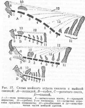
U pigs The cervical spine is very short (D), strong and relatively little mobile, especially in the lateral directions. The spinous processes protrude strongly on it. The transverse costal processes are equipped with wide plates directed downward, overlapping one another in a tiled manner. This construction shows that the neck as a short lever is used mainly for extension and flexion and is in connection with the way these animals in the wild obtain nutritious material, which they extract from the soil, rummaging in it with a short proboscis. In pigs, in addition to extremely well-developed muscles that extend and flex the cervical lever and the head (in the occipito-atlas joint), there are also significant lateral cervical muscles, since the roots dug by the trunk are pulled out with a sideways curved canine.
U cattle cervical region spinal column relatively short (C). It is still relatively longer than that of pigs, but shorter than that of horses. The head and neck are easily supported by the powerful nuchal ligament (12, 13). The spinous processes clearly protrude. The transverse costal processes give off lamellar shoots from the sides downwards, which, however, are less developed than in pigs. The muscles acting on the cervical lever in cattle also have a specific purpose - to strongly and quickly raise the lowered head upward in order to use the horns, a weapon of self-defense. Therefore, he has highly developed dorsal neck muscles. A cross-section of the neck as a whole, with muscles and skin, shows an oval, or more precisely ovoid, shape with a pointed upper end and a blunt lower end.
U horses the neck lever can be considered the longest compared to that of previous types (4). The neck and head are held in a resting hanging position without any expenditure of muscular strength by the extremely well-developed nuchal ligament (12, 13). The spinous processes on the cervical vertebrae, with the exception of the 7th, are completely undeveloped. The transverse costal processes do not give rise to pronounced lamellar projections downwards, with the exception of the 6th vertebra. The transverse section of the neck generally approaches an ovoid shape with a blunt edge directed ventrally and a sharp edge dorsally. The neck bends relatively easily in all directions, especially in the midsagittal plane. It should be noted that the length and mobility of the neck of horses varies among various breeds; Heavy walking horses have a shorter neck, less mobile (pig's neck), while fast horses have a longer and more agile neck. This is understandable, since the neck with the head suspended serves as a good balancing device during fast gaits when removing the center of gravity of the body from stable balance.
The first reptiles appeared on Earth several hundred million years ago. Their high level of organization allowed them to easily destroy invertebrate competitors, becoming sole “rulers.” From that time on their heyday began. The dominance of reptiles on our planet has reached unimaginable proportions! They not only captured all the land, but also returned to the water, where they significantly displaced the ancient fish that reigned there. Moreover, the reptiles rose into the air! Today, reptiles are one of the most interesting, diverse and bright groups animals on planet Earth. It is interesting that people do not have such a contradictory attitude towards any class of animals as they do towards reptiles: they are loved and at the same time hated, they evoke childish interest and mystical horror. Externally, reptiles can vary significantly. Next, we will consider several types of their structure.
Lizard, snake and tortoiseshell types of reptile structure
Perhaps the most common structure of reptiles is the so-called lizard type. Individuals belonging to it have a head, a cervical region, a torso itself, a tail and four limbs. These are lizards, crocodiles, tuataria. The next type is snake. There’s probably no need to explain that limbs are simply out of the question for representatives of this appearance! These are flexible reptiles with an elongated body - snakes and many lizards that have lost their limbs. Representatives of the tortoiseshell type of structure, in all respects, fit the lizard structure, but only with one more feature - a body enclosed in a powerful shell. Of course, these are turtles. 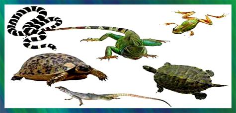 Now let's talk about the characteristics that distinguish reptiles not within their own group, but, so to speak, on the “world stage.”
Clad in "armor"
The main characteristics of reptiles that distinguish them from other animals are, of course, the peculiarities of movement on the ground and leather “armor”. We have already talked about the first feature. Let's look at the second sign. The fact is that a powerful stratum corneum is developed in the skin of reptiles, which forms scutes and scales. Almost the entire body of the animal is covered with this “armor”, even the eyelids and the thin skin between the fingers! This leather “chain mail” is a very durable protection, and the shields that usually cover the upper part of the head are a kind of “helmet”. Sometimes bone plates grow under the horny scutes. They, like armor, protect the body of a reptile. 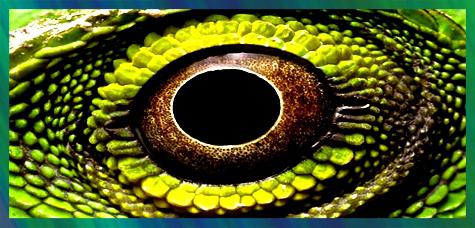 This is how crocodiles, lizards, snakes and other scaly land animals are arranged in an interesting way. But what about their flexibility? Does this “chain mail” really prevent them from even turning their heads? Fortunately, this is not the case! Why do reptiles need head mobility? The ability to turn your head plays a big role in reptile hunting and safety. Let's talk about this in more detail.
Why do reptiles need head mobility?
As you know, reptiles are a continuation of amphibians that came to land. One of the features that distinguishes reptiles from amphibians is the presence of a cervical spine. For clarity, let's say that, for example, frogs do not have a neck, and the head immediately goes into the body. The neck allows the reptile to turn its head to the side. You may ask why do reptiles need head mobility? We answer with an example. Having heard some sound or rustle of a moving object, the lizard instantly turns its head in its direction and sees everything that is happening. If it is in danger, it runs away, but if it is potential prey, then the lizard grabs it, quite skillfully dealing with it. The ability to turn their heads in different directions makes reptiles flexible, almost elusive, and also lightning-fast hunters. This is why reptiles need head mobility.
Interesting things about snakes
Perhaps the most unique reptiles are snakes. They are adapted to crawl without the help of limbs and swallow large prey whole. Snakes are fundamentally different from other reptiles in that they have a very long and elastic body, which, in turn, leads to a very peculiar arrangement internal organs. 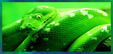 As we know, in ordinary vertebrates many organs are paired and arranged symmetrically. In snakes, only half of them almost always remains; for example, many of them have only the right lung. And if there are paired organs, then they are located at different ends of the long body. For example, the right kidney is shifted towards the head, the left - towards the tail! When we hear the word “snake,” thoughts of a poisonous reptile involuntarily come to mind. But today science knows more than 2,700 and only 700 of them are poisonous.
1. Write an answer about the cell membrane.
The shell not only delimited the contents of the cell from the external environment, but also allowed it to increase the speed of movement due to flagella and cilia.
2. Let's write an answer about the support function.
Yes. The covers of worms perform support function, since they limit the contents of the body, maintain the shape of the body, since they are quite strong and dense (for example, the cuticle of roundworms).
3. Let us explain the importance of the chitinous shell.
Muscles are attached to the chitinous shell of arthropods, which allows the animals to move (running, jumping, flying) and the ability to quickly spread.
4. Let us define the concept.
Joint.
5. Let us indicate the first animal with a chord.
Lancelet.
Let us define the concepts.
The chord is a long elastic longitudinal cord inherent in representatives of the chordate type.
A vertebra is a constituent element of the spinal column.
The sacrum is a large, triangular-shaped bone located at the base of the spine that forms the upper back the pelvic cavity, like a wedge, located between the two pelvic bones.
6. Let's label the parts of the vertebra.
1 – vertebral body
2 – upper arcs
3 – lower arches
4 – canal of the spinal cord.
7. Let's write about the influence of the aquatic-terrestrial lifestyle.
The axial skeleton has become more complex. The cervical region (1 vertebra) and the sacral region (1 vertebra) appeared; the trunk region was represented by 7 vertebrae with ribs. Tailed animals have caudal section(several vertebrae).
8. Let's write an answer about head mobility.
Head mobility is a necessary condition for terrestrial existence.
9. Let us explain why birds need light bones.
The lungs and hollow inside of bird bones are a device for flight.
10. Let's distribute the parts of the skeleton.
Skeleton parts:
1. Thigh.
2. Shoulder.
3. Shin.
4. Brush.
5. Forearm.
Answer:
A – 2, 4, 5.
B – 1, 3.
11. Let's write an answer about the appearance of the sternum and chest.
The sternum and rib cage first appeared in reptiles. These parts of the skeleton protect the heart and lungs and provide better oxygen supply to the lungs.
12. Let's write an answer about swallowing large prey.
In snakes, the ends of the ribs swing freely.
13. Let's make diagrams.
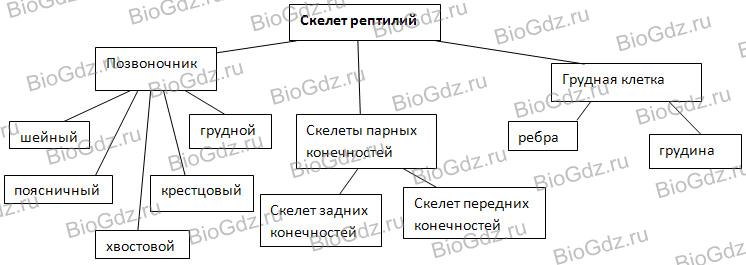
Differences: reptiles have new sections of the spine: lumbar and sacral instead of the trunk. The tail is well developed. The chest with ribs appears. In the cervical region the vertebrae are connected movably.
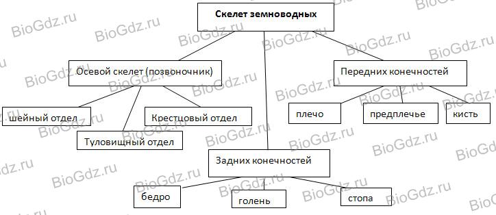
14. Let's make a diagram of the skeleton of mammals.
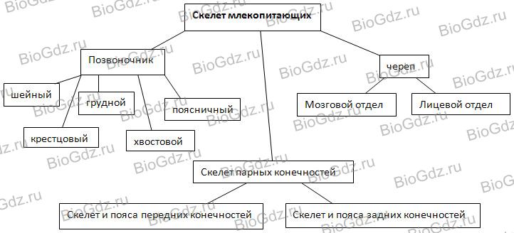
15. Let's write about the complications of the skeletal structure.
Mammals have conquered all habitats: land, air, water; began to move faster. They have on high level The respiratory, circulatory and other organ systems are developed, so improvement of the skeleton is a regularity and a necessary condition for the existence of mammals.
Anatomy, morphology and ecology of reptiles
4. Reptile skeleton
The vertebral column of reptiles is divided into five departments: cervical, thoracic, lumbar, sacral and caudal.
Education flexible neck and gain head mobility was of paramount importance in obtaining food and in orientation. The mobility of the head is ensured by the differentiation of the first two cervical vertebrae - the atlas, or atlas (atlas), and epistropheus (epistropheus). The atlas has the appearance of a bone ring, divided by a dense ligament into upper and lower halves; through the superior opening the brain is connected to the spinal cord; the anterior surface of the lower half articulates with the occipital condyle of the skull, and at the back the odontoid process of the second cervical vertebra, the epistropheus, enters the lower foramen. The head may turn to the sides on the odontoid process, and its movement in the vertical plane is ensured by the articulation of the cranial condyle with the atlas. All this ensures complex movements of the head, enhanced by the mobility of the entire neck. The study of embryonic development showed that the odontoid process is formed by accretion of the body of the atlas to the epistrophy.
Neck movements are determined by number and structure vertebrae cervical region; they are different in different groups. In the hatteria, the vertebrae are still amphicoelous (fish-type) with remnants of the notochord between them. In crocodiles and most squamate vertebrae are procoelous (anteroconcave) and only in a few lower forms are amphicoelous. Some of the cervical vertebrae bear short ribs. In cryptonecked turtles, which bend their necks in a vertical plane, the cervical vertebrae retain only the rudiments of transverse processes. On the contrary, in side-necked turtles, which bend their necks to the side, the transverse processes and associated muscles are highly developed. The complex movements of the neck of turtles are also ensured by the variety of vertebrae: the posterior vertebrae are procoelous, the anterior ones are opisthocoelous (retroconcave), and the middle one is amphicoelous.
TO thoracic vertebrae long ones are articulated ribs , the abdominal ends of which are attached to the sternum , forming a closed chest (snakes do not have a chest). The shoulder girdle is also attached to the sternum. The lumbar vertebrae also bear ribs that do not reach the sternum. TO sacral region, consisting of two vertebrae, is attached pelvic girdle . The caudal region helps maintain balance when moving, and sometimes serves as propulsion (in aquatic snakes, crocodiles and some aquatic lizards).
In lizards capable of autotomy (tail drop), the caudal vertebrae can break in the middle, where there are thin cartilaginous layers dividing the vertebral body into two parts.
Total number vertebrae varies in different species and reaches 50-80 (7-10 cervical, 16-25 sternolumbar, 2 sacral, 15-40 caudal). In snakes and legless lizards, the number of vertebrae increases, and the spine is divided only into the trunk and caudal sections. All trunk vertebrae are equipped with movable ribs that rest against the ventral scutes, which is important when snake-like movement. The total number of vertebrae increases to 140 (in thick and short snakes) - 435 (in snakes with a long body).
Paired limbs and their belts
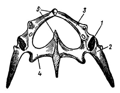
Shoulder girdle reptiles is similar to the amphibian belt, but it has highly developed ossification. Coracoid (coracoideum) at the junction with spatula (scapula) form the articular cavity for the articulation of the head of the humerus (Fig. 3).
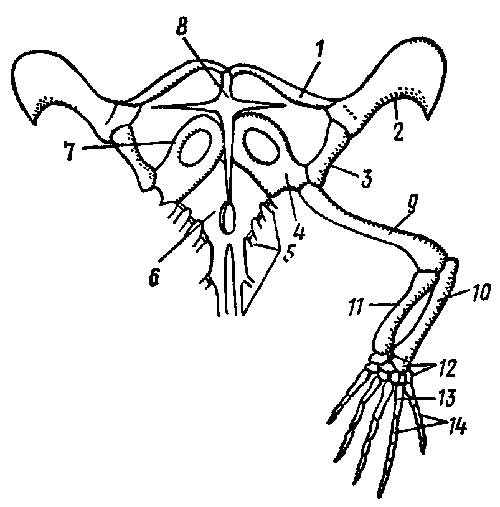
Rice. 3. Shoulder girdle and forelimb of the Lacerta lizard: 1 - clavicle, 2 - suprascapular cartilage, 3 - scapula, 4 - coracoid, 5 - ribs, 6. - sternum, 7 - procoracoid cartilage, 8 - episternal, 9 - shoulder, 10 - ulna, 11 - radius, 12 - wrist, 13 - metacarpus, 14 - phalanges
A flattened suprascapular cartilage (cartilage suprascapularis), and in front of the coracoid - cartilaginous procoracoid (cart. procoracoidea). The coracoid and procoracoid of each side are fused with the unpaired bone sternum (sternum); Through the chest, the girdle of the forelimbs is attached to the axial skeleton. From below, a cruciform integumentary bone grows to the sternum - episternum (episternum). Paired coverslips collarbone (clavicula) connect the anterior end of the episternum to the dorsal part of each scapula.
This design enhances strength shoulder girdle. In turtles, the episternum and clavicles are included in abdominal shield shell; in crocodiles only the coracoids and scapulae are well developed.
Pelvic girdle consists of two nameless bones; each of them is formed by the merger of three pelvic bones - ileal (ilium), ischial (ischium) and pubic (pubis), together forming the acetabulum, which forms a joint with the head of the femur (Fig. 14).
Rice. 4. Pelvic girdle of the Lacerta lizard: 1 - articular cavity for the femoral head, 2 - ilium, 3 - pubis, 4 - ischium, 5 - symphysis
The ilia articulate with the transverse processes of the sacral vertebrae. All modern reptiles have a pelvis closed: the right and left pubic and ischial bones are connected to each other along midline symphysis - cartilaginous bridge.
Doubles limbs differ in different species and groups of reptiles depending on the predominance of certain methods of movement. But they usually retain the general structure of the paired limbs of terrestrial vertebrates.
Unlike amphibians, in reptiles the movable joint in the forelimb is located between two rows of carpal bones ( intercarpal joint), and in the hind limb - between the rows of tarsal bones ( intertarsal joint).
Reptile skull modified mainly depending on the nature of nutrition and methods of obtaining food. It differs from the skull of amphibians in its elongated jaws, forming a relatively long snout ; the wide and short skull of amphibians was necessary for their oropharyngeal breathing mechanism; at the same time, the wide mouth contributed to the capture of small prey when thrown at it. In reptiles, breathing is ensured by work chest, and the capture of prey is associated with active pursuit, in which the elongated snout has an advantage. This shape of the jaws also made it possible to tear off pieces from large prey. Both required more powerful chewing muscles. The main changes in the skull of reptiles are associated with its development and the complication of the sense organs.
The skull is almost completely ossified (Fig. 5).
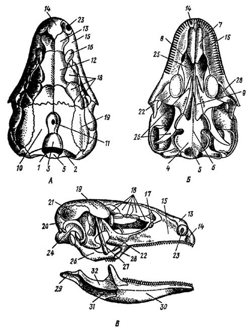
Rice. 5. Lacerta lizard skull(according to Parker). A - top view; B - bottom view, B - side view
1 - foramen magnum, 2 - lateral occipital bone, 3 - superior occipital bone, 4 - main occipital bone, 5 - occipital condyle, 6 - main sphenoid bone, 7 - vomer, 8 - choana, 9 - parasphenoid, 10 - parietal bone, 11 - interparietal bone with an opening for the parietal organ, 12 - frontal bone, 13 - nasal bone, 14 - premaxillary bone, 15 - maxillary bone, 16 - prefrontal bone, 17 - lacrimal bone, 18 - supraorbital bones, 19 - postfrontal, or postorbital bone, 20 - squamosal, 21 - supratemporal bone, 22 - zygomatic bone, 23 - nostril, 24 - quadrate bone, 25 - palatine bone, 26 - pterygoid bone, 27 - upper pterygoid, or columnar bone, 28 - transverse bone, 29 - articular bone, 30 - dentary, 31 - surangular bone, 32 - coronoid bone
The occipital region consists of four occipital bones (occipitalia) of chondral origin: superoccipital, main and two lateral. They border the foramen magnum, below which lies the only occipital condyle , formed by the main and both lateral occipital bones. Integument main sphenoid bone (basisphenoideum) lies in front of the main occipital, forming the bottom of the skull. In front of it grows a small parasphenoid (parasphenoideum) and paired openers (vomer), on the side of which lie choanae . In the area of the auditory capsule there are three ear bones (otici); anterior ear, maintaining independence; posterior auricular, fused with the lateral occipital, and superior auricular, fused with the superoccipital. There are no bones in the olfactory region; it remains cartilaginous.
Skull roof formed by paired integumentary bones: nasal (nasalia), prefrontal (praefrontalia), frontal (frontalia) and postfrontal (postfrontalia); further lie parietal (parietalia) and unpaired interparietal (interparietale) bones; the latter has an opening for the parietal organ.
Sides of the skull form integumentary bones: paired intermaxillary (intermaxillare) in some species merging, paired maxillary (maxillare), supraorbital (supraorbitale), zygomatic (jugale), quadratozygomatic (quadratojugale) (the quadratojugal bone is present in the anapsid and typical diapsid skulls of hatterias and crocodiles, but disappears in lizards that have a diapsid type of skull with a reduced lower arch) and scaly (squamosum). Palatoquadrate cartilage in the posterior part it gives rise to paired chondral ossifications - square bones (quadratum), the upper part is connected to the braincase, and the lower part is connected to the lower jaw.
The anterior part of the palatoquadrate cartilage is replaced by integumentary bones, forming bottom of the skull: paired palatal (palatini) and pterygoid (pterygoidei). Transverse bones (transversi) connect the pterygoid bones with the maxillary bones, and in lizards and tuataria also superior pterygoid , or columnar (epipterygoidei), bones connect the pterygoid bones with the parietal bones.
In turtles and especially in crocodiles, the growth of the palatine processes of the premaxillary and maxillary bones, as well as the palatine bones, forms secondary bony palate dividing the oral cavity into the upper section - nasopharyngeal , and the bottom one is actually oral cavity . Therefore, the choanae move back towards the larynx, which allows breathe when only the end of the head with the nostrils is exposed from the water.


