The tail section of the spine in reptiles. Lizard skeleton. The internal structure of the lizard. Types and names of lizards
Vertebral column reptiles are divided into five sections: cervical, thoracic, lumbar, sacral and caudal.
The formation of a flexible neck and increased mobility of the head were of paramount importance in obtaining food and in orientation. The mobility of the head is provided by the differentiation of the first two cervical vertebrae - the atlas, or atlas (atlas), and the epistropheus (epistropheus). The atlas looks like a bony ring divided by a dense ligament into the upper and lower halves; through the upper opening, the brain connects to the spinal cord; the anterior surface of the lower half articulates with the occipital condyle of the skull, and the dentate process of the second cervical vertebra, epistrophy, enters into the lower foramen behind. The head can be rotated to the sides on the odontoid process, and its movement in the vertical plane is provided by the articulation of the cranial condyle with the atlas. All this provides complex head movements, enhanced by the mobility of the entire neck. The study of embryonic development showed that the odontoid process is formed by accretion to the epistrophy of the atlas body.
Neck movements are determined by the number and structure of the cervical vertebrae; they are different in different groups... In the tuatara, the vertebrae are still amphitic (fish type) with remnants of a notochord between them. In crocodiles and most of the scaly vertebrae, the vertebrae are procelial (antero-concave) and only in a few lower forms are amphitic. Part of the cervical vertebrae bears short ribs. In latent-cervical turtles, bending the neck in a vertical plane, the cervical vertebrae retain only the rudiments of the transverse processes. On the contrary, in side-necked turtles, bending the neck to the side, the transverse processes and the muscles associated with them are highly developed. The complex movements of the turtles' neck are also provided by a variety of vertebrae: the posterior vertebrae are procelian, the anterior vertebrae are opistocoelous (posterior concave), and the middle vertebrae are amphitic.
TO thoracic vertebrae long ribs are attached, the abdominal ends of which are attached to the sternum with the help of cartilaginous sections, forming a closed chest ( chest no snakes). The shoulder girdle is also attached to the sternum (see below). The lumbar vertebrae also bear ribs that do not extend to the sternum. The pelvic girdle is attached to the sacral region, which consists of two vertebrae. The tail section helps to maintain balance during movement, and sometimes serves as a mover (in water snakes, crocodiles and some water lizards). In lizards capable of autotomy, the caudal vertebrae can fracture in the middle, where there are thin cartilaginous layers that divide the vertebral body in two.
Axial skeleton... The differentiation of the axial skeleton, or spine, into divisions is expressed in reptiles much more clearly than in amphibians. The cervical spine is always composed of several vertebrae, of which the two anterior ones have a special structure. The first cervical vertebra is called atlas or atlas. It is devoid of a vertebral body and has the form of a ring divided into two parts. On the lower front surface of this vertebra there is an articular cavity, which is movably connected to the condyle of the skull. The second cervical vertebra - epistrophy, has a large odontoid process in front, which is the body of the first cervical vertebra, fused with the epistrophy. The dentate process freely enters the lower opening of the atlas. This structure of the first cervical vertebrae provides greater mobility of the head. The rest of the cervical vertebrae have the usual structure; many of them have short cervical ribs.
.
A - atlas; B - epistrophy; B - thoracic vertebra;
D - longitudinal section of the thoracic vertebra:
1 - odontoid process of the epistropheus, 2 - vertebral body, 3 - upper arch,
4 - spinous process, 5 - canal for the spinal cord, 6 - anterior articular process,
7 - posterior articular process
The thoracic and lumbar regions are not clearly distinguished and are usually considered as a single department. The actual thoracic region is that part of the spine in which the ribs extending from the vertebrae are attached to the sternum with their lower end. Vertebrae lumbar have ribs that do not reach the sternum. The vertebral bodies are concave in front and convex in the back; such vertebrae are called procellular. The upper arches rise above the vertebral body, ending in a spinous process. The spinal cord is located in the canal formed by the upper arches.
From the anterior and posterior parts of the base of the upper arches, the anterior and posterior articular processes depart, respectively. These paired processes connect with the articular processes of adjacent vertebrae and contribute to the greater strength of the spine when bending. On the sides of the vertebral body (near the base of the upper arches) there are small depressions to which the ribs are attached.
The sacral region consists of two vertebrae, which are characterized by powerfully developed transverse processes; they are joined by the bones of the pelvis. The caudal region is represented by numerous vertebrae, gradually decreasing in size.
This structure of the spine is typical for the class of reptiles, but in some groups it undergoes secondary changes. In particular, in snakes, due to the reduction of paired limbs and the emergence of a different type of movement - crawling on the belly by bending the body - the spine is clearly divided only into the trunk and tail sections. All trunk vertebrae have movable ribs, the lower ends of which are free (the sternum is absent in snakes) and abut against the abdominal horny shields.

.
A - carapace; B - plastron:
1 - trunk section of the spinal column, 2 - costal plates,
3 - marginal plates, 4 - coracoid, 5 - scapula, 6 - ilium,
7 - pubic bone, 8 - ischium
In turtles, the axial skeleton takes part in the formation of the bone base of their shell. The upper shield of the carapace - carapace - is composed of several rows of bone plates. The middle (unpaired) row of these plates is formed by the fusion of expanded and flattened spinous and transverse processes of the trunk vertebrae with skin bones; on the sides of the middle row are paired rows of bone plates fused with extended ribs. The edge of the carapace is formed by bony plates of integumentary origin. Thus, the torso spine of turtles is motionless and firmly adhered to the dorsal shield of the shell. The cervical and tail sections of the spine are mobile. In this case, the anterior cervical vertebrae are opisthocoelous (the vertebral body is convex in front, concave in the back), the posterior ones are longitudinal, and between these two groups there is one vertebra, the body of which has a convex surface both in front and behind.
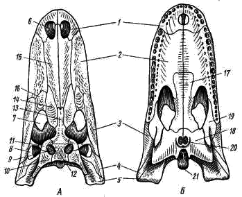
Scull... Compared with amphibians, the skull of reptiles is characterized by a much more complete ossification. A certain amount of cartilage is preserved only in the olfactory capsule and in the auditory region. The axial and visceral parts of the skull are embryonic laid separately, but in adult animals they are unified education... The skull includes both cartilaginous (replacement, or primary) and numerous skin (integumentary, or secondary) bones. It is convenient to use the skull of a large lizard, the monitor lizard, as the main object for study.

.
A - on the side; B - from below; B - from above; G - behind:
1 - the main occipital bone, 2 - the lateral occipital bone,
3 - superior occipital bone, 4 - large occipital foramen,
5 - occipital condyle, 6 - anterior auricular bone,
7 - main sphenoid bone, 8 - opener, 9 - parietal bone,
10 - frontal bone, 11 - nasal bone, 12 - prefrontal bone,
13 - preorbital bone, 14 - lacrimal bone, 15 - superior temporal fossa,
16 - posterior bone, 17 - squamous bone, 18 - premaxillary bone,
19 - maxillary bone, 20 - zygomatic bone, 21 - rupture of the lower temporal arch due to reduction of the square-zygomatic bone, 22 - square bone,
23 - pterygoid bone, 24 - palatine bone, 25 - upper pterygoid bone,
26 - transverse bone, 27 - supracular bone, 28 - dentary, 29 - angular bone,
30 - articular bone, 31 - coronoid bone
Axial skull
... In the occipital region of the skull, there are all four occipital bones: the main occipital, two lateral occipital, and the superior occipital. These primary bones surround the foramen magnum. The lower and lateral occipital bones together form a single (in contrast to amphibians) occipital condyle, movably articulating with the first cervical vertebra - atlas. The articulation of the head with the neck with the help of only one condyle, in combination with the already discussed structural features of the first two cervical vertebrae, gives the head of reptiles considerable mobility.
In the auditory section, of the cartilaginous bones, only the paired anterior auricular bone retains its independence, while the superior auricular fuses with the superior occipital bone, and the posterior auricular - with the lateral occipital.
The interorbital septum in reptiles is thin, membranous, and only crocodiles and lizards have separate small ossifications in it, apparently corresponding to the eye-wedge-shaped bones. The olfactory capsule has no ossification.
At the base of the skull, in front of the main occipital bone, there is a rather large integumentary main sphenoid bone. Its anterior narrow process is homologous to the parasphenoideum, which is markedly reduced in reptiles. In the front part of the bottom of the skull, under the olfactory section, there is a paired opener, which also has an integumentary origin.
The roof of the skull is represented by numerous integumentary bones, some of which descend from top to bottom and cover the skull from the sides. These include the parietal, frontal, and nasal bones. In front of the frontal bones, paired prefrontal and preorbital bones are usually located, and under them in the anterior wall of the orbit are paired lacrimal bones perforated by a narrow canal.
A - from above; B - from below:
1 - premaxillary bone, 2 - maxillary bone, 3 - zygomatic bone,
4 - square-zygomatic bone, 5 - square bone, 6 - external nostril,
7 - orbit, 8 - lateral temporal fossa, 9 - superior temporal fossa, 10 - squamous bone,
11 - posterior frontal (postorbital) bone, 12 - parietal bone, 13 - frontal bone,
14 - prefrontal bone, 15 - nasal bone, 16 - lacrimal bone, 17 - palatine bone,
18 - pterygoid bone, 19 - transverse bone,
20 - choanas (internal openings of the nostrils), 21 - occipital condyle
Of the rest of the integumentary bones of the axial skull, bones that take part in the formation of the so-called temporal arches are of particular interest. In a crocodile, in the roof of the skull, outward from the parietal bone, on each side there is an opening - the upper temporal fossa. Along the outer edge, the superior temporal fossa is bounded by the posterior frontal or postorbital ridge bones. These two bones together make up the superior temporal arch. On the side of the skull, behind the orbit, there are lateral temporal fossae, bounded from the outside by the lower temporal arches. Each lower temporal arch is composed of two bones: the zygomatic and the square-zygomatic. The lower temporal arch is connected to the upper jaw: the zygomatic bone grows to the maxillary, and the square-zygomatic bone grows to the square. This type of skull, like that of a crocodile - with two temporal pits and two temporal arches, is called diapsid (two-arc).
In the monitor lizard, the upper temporal fossa is limited by the complete upper temporal arch, while in the lower temporal arch, the square-zygomatic bone was reduced and only the zygomatic bone was preserved; the lateral temporal fossae are therefore not closed from the outside and remain open. Therefore, the skull of a monitor lizard can be considered as a skull of the diapsid type, but with a reduced lower arch. In some other lizards, the upper temporal arch is also partially reduced, and in snakes both temporal arches are reduced (the posterior forehead and squamous bones do not connect to each other; both temporal fossae remain open from the outside). Thus, snakes and lizards (squamous order, Squamata), according to the structure of the skull, belong to the group of diapsid (two-arc) reptiles, but are characterized by a different degree of reduction of the temporal arches.
:
1 - false temporal fossa, 2 - premaxillary bone, 3 - maxillary bone,
4 - zygomatic bone, 5 - square-zygomatic bone, 6 - square bone,
7 - scaly bone, 8 - posterior bone, 9 - parietal bone,
10 - frontal bone, 11 - prefrontal bone, 12 - superior occipital bone
In the tortoise, both temporal pits are absent, and the lateral wall of the skull roof, delimiting a large cavity from the outside - the so-called false temporal fossa, formed as a notch in the occipital part of the skull, is composed of densely fused bones: posterior forehead, scaly, zygomatic and square-zygomatic. This type of skull, devoid of true temporal pits and the temporal arches limiting them, is called anapsid (arcless).
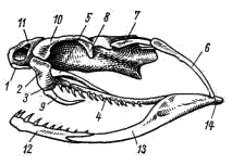
Visceral skull ... In the monitor lizard, the palatine-square cartilage ossifies, forming a square bone in the posterior part, to the lower end of which it adjoins lower jaw; the upper end of the square bone is movably articulated with the axial skull. In front of the square bone is the pterygoid, and in front of it is the palatine bone, which connects to the maxillary bones and the vomer. All these bones are paired; of these, only square bones are of cartilaginous (primary) origin.
The upper pterygoid bone extends upward from the pterygoid bone. This paired bone, connecting the pterygoid and parietal bones, is homologous to the vertical (“ascending”) process of the palatine-square cartilage and is characteristic of living reptiles for lizards and tuatara. In addition to the upper pterygoids, the transverse bones extend from the pterygoid bones, which in their front part join the maxillary bones. Secondary upper jaw represented by the premaxillary and maxillary bones. The lower jaw consists of the primary articular bone and integumentary bones: dental, angular, supra-angular, coronal and, sometimes, several more small bones.
On the premaxillary, jaw and dental bones of reptiles (except for turtles) there are simple conical, sometimes slightly bent back teeth, which grow to the edge of the corresponding bone.
The hypoglossal arch, like in amphibians, has completely lost the function of a suspension. The upper element of the hyoid arch (hyomandibular) is part of the middle ear in the form of a rod-shaped auditory ossicle (stapesseucolumella), and the rest of it, together with the remnants of the anterior branchial arches, forms the hyoid apparatus.
The described structure of the visceral skull is generally typical of all reptiles. But in some groups there are deviations from this scheme, mainly associated with the specifics of the biology of these groups.
:
1 - premaxillary bone, 2 - maxillary bone. 3 - palatine bone,
4 - pterygoid bone, 5 - transverse bone, 6 - square bone, 7 - squamous bone,
8 - posterior bone, 9 - poisonous tooth, 10 - frontal bone, 11 - nasal bone,
12 - dental bone. 13 - angular bone, 14 - articular bone
In snakes, not only square bones are very mobile, but also scaly bones connected to them, as well as pterygoid and palatine bones. The last two have sharp teeth. The transverse bones in snakes serve as levers, transmitting the movements of the pterygoids to the maxillary bones, which in turn are very mobile. This entire system of movably articulated bones not only contributes to an extremely wide opening of the mouth, but also provides independent movements of the right and left halves of the jaw apparatus when pushing prey into the pharynx with alternate interception. This allows snakes to swallow relatively very large (exceeding the thickness of the snake's body) prey. Have poisonous snakes on the maxillary bones there are movably attached sharp, bent back poisonous teeth, which have an internal canal or groove on the front surface, along which, when bitten into a wound, poison flows from the poisonous glands located at the base of the tooth.
The crocodile skull is characterized by the fact that the teeth do not adhere to the edge of the dental, premaxillary and maxillary bones, as in other reptiles, but sit in special depressions (holes, or alveoli) of these bones - thecodont teeth. Another feature of the visceral skull of crocodiles is the secondary hard palate, which separates oral cavity from the nasopharyngeal passage. The palatine processes of the premaxillary and maxillary bones, as well as the palatine and pterygoid bones, take part in the formation of the secondary hard palate. Due to the formation of a hard palate, the secondary choans are carried back and are located in the pterygoid bones, above the larynx. The formation of a secondary hard palate is associated with the nature of the crocodiles' lifestyle: direct contact of the larynx with the choans opens up the possibility of uninterrupted breathing when eating and when the crocodile rests in shallow water, exposing the nostrils on elevations from the water, while the oral cavity is filled with water.
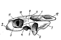
Paired limbs and their belts. Shoulder girdle reptiles consists of typical bones: the scapula located more dorsally and the coracoid facing the ventral side. Both of these bones are involved in the formation of the glenoid fossa for the attachment of the forelimb. Dorsal to the scapula, there is a wide, flattened suprascapular cartilage, and in front of the coracoid there is a cartilaginous procoracoid. There is a well-developed sternum, to which several ribs are attached. Thus, unlike amphibians, reptiles develop a thorax and the shoulder girdle is supported in the axial skeleton. On the ventral side of the sternum, there is a T-shaped integumentary bone - the supra-sternum, in front of it - also integumentary bones - the clavicle. The outer ends of the collarbones are attached to the shoulder blades, and the inner ends are fused with the branches of the episternum. The collarbone and the episternum (absent in amphibians) increase the strength of the connection between the right and left sides of the shoulder girdle.

Lizard's shoulder girdle (bottom view):
1 - scapula, 2 - suprascapular cartilage, 3 - coracoid,
4 - glenoid cavity for the shoulder head, 5 - procoracoid cartilage,
6 - sternum, 7 - ribs, 8 - brisket, 9 - clavicle
In snakes, the shoulder girdle is completely reduced, and in turtles, the clavicle and the episternum became part of the bones of the abdominal shield of the carapace, forming, respectively, the anterior paired and unpaired bone plates wedged between them.

Lizard's pelvic girdle (bottom view):
1 - ilium, 2 - pubic bone, 3 - ischium,
4 - acetabulum (articular fossa) for the femoral head,
5 - sacral vertebrae
The pelvic girdle consists of two symmetrical halves connected by midline cartilage. Each half is made up of three bones; located dorsally iliac, located on the ventral side of the pubic and sciatic. All these bones take part in the formation of the articular fossa, to which the hind limb is attached. The reptile's pelvis is closed: the right and left pubic and ischial bones on the ventral side are fused together.

.
A - front; B - back:
1 - brachial bone, 2 - ulna, 3 - radius, 4 - wrist,
5 - metacarpus, 6 - phalanges of fingers, 7 - intercarpal joint, 8 - femur,
9 - tibia, 10 - fibula, 11 - knee cap,
12 - tarsus, 13 - intertarsal joint, 14 - metatarsus
The limbs of reptiles are built according to the typical pattern of limbs of terrestrial vertebrates. The proximal part of the forelimb is represented by one bone - the humerus, followed by the forearm, which consists of two bones - the ulna and the radius. The wrist consists of relatively small bones, usually arranged in two rows; on the side of them is another pear-shaped bone, taken for the remainder of the sixth finger. The metacarpus is composed of five elongated bones, to which the phalanges of five fingers are attached. The last phalanxes bear claws. The joint that provides mobility of the hand in reptiles does not pass between the bones of the forearm and the proximal row of the bones of the wrist (as in amphibians), but between the proximal and distal rows of the bones of the wrist. This joint is called intercarpal joint.
In the hind limb, the proximal element - the thigh articulates knee joint with a tibia, consisting of two tibia bones - large and small. Above the front surface of this joint is a small bone called the patella. In the tarsus, the proximal row of bones grows together or almost motionlessly connects with the bones of the lower leg, and the bones of the distal row are also closely connected and partially fused with the metatarsal bones. Due to this, the articular surface is located here not between the tibia and the foot, but between the proximal and distal rows of the tarsal bones. Such a joint is typical for reptiles and is called the intertarsal joint. The metatarsus consists of five elongated bones, to which the phalanges of five fingers are attached. The terminal phalanges bear claws.
Body shape reptiles are much more variable in their manifestations, in comparison with amphibians, which is explained by their habitation in more diverse ecological conditions and a variety of modes of movement.
Lizardlike the shape of reptiles is characteristic of many species (lizards, chameleons, crocodiles) and is determined by movement over the substrate with support on both pairs of limbs. These reptiles have more distinctly parts of the body are highlighted - head, neck, torso and tail.
Limbs, providing movement, are located, like amphibians, on the sides of the body... This position of the limbs and their small length does not contribute to the rapid movement of animals with a large body weight (crocodiles). However, for most species this is not a hindrance - reptiles run fast, including on uneven and vertical surfaces.
Snakes and legless lizards, moving by bending the body, acquired a different shape - sharply elongated, without clear differentiation of the body into sections. This way of transferring the body ("crawling"), which provides good adhesion to the surface, allows you to successfully move in different conditions - on the ground, in the soil, in the water and on trees.
In reptiles, for the most part living in aquatic environment(crocodiles, sea snakes and turtles), the shape of the body, in many ways, as in terrestrial conditions, is determined by the way of movement. Torso crocodiles somewhat flattened in the dorso-ventral direction, while the tail, which serves as a rudder, is sharply flattened laterally; between the toes of the hind limbs there is swimming membrane... The tail section has the same shape. at sea snakes with a rounded cross-section of the body. Sea turtles characterized by flattening and expansion of the shell, which determines the body's support on water, and flipper limbs.
The main manifestations external signs reptiles are well traced on example of lizards.
In general outlines, they are very similar to tailed amphibians (salamander, newt) - body elongated, with a rounded section and support on both pairs of limbs. Head most lizards are pointed anteriorly and flattened from top to bottom, covered with rather large scutes, the shape and location of which, like on the body, is of a specific nature.
On the front of the head there is a rather wide mouth opening... On the sides of the head are located eyes with movable eyelids and blinking membrane, and in front of them - paired nostrils.
In the back of the side of the head lie large auditory holes tightened eardrum, which is located in a small depression of the skin, as a result of which outer ear bud... Near the occiput, in the center of the inter-parietal scutellum is noticeable vestigial parietal eye.
On the border trunk and tail departments visible cloacal gap, which has a transverse arrangement (lizards, snakes) and a longitudinal arrangement in turtles and crocodiles.
Body covers reptiles consist of two layers - multilayer epidermis and corium. The epidermal layer is heterogeneous: its upper layer is constantly keratinized, due to the filling of cells with grains of a special protein - keratin. As a result, cells lose their characteristic structure (protoplasm, nucleus) and die off. Below lies another layer - malpighiev consisting of living, constantly dividing cells.
The upper layer of the epidermis serves as the basis for the formation of horny formations - scales, scutes, thorns, claws... In some species of reptiles ( crocodiles) larger formations develop under them - bone plates, or osteoderm.
Under the epidermis of turtles, bone-horny or bone-leathery forms carapace, which serves as a passive defense against enemies. It consists of a top (carapace) and lower ( plastron), which are connected by a leathery or bone jumper. The spine and girdles of the limbs take part in the formation of the shell.
In general, reptile skin is relatively thin, dry and glandless... Only in species of individual taxonomic groups do unicellular glands located on different parts of the body. In lizards, they are in the area of the cloaca and on inside hips ( thigh pores), others (snakes, turtles, crocodiles) on the head and other places. The secretions of these glands perform information function: used to mark an individual territory and serve to attract individuals of the opposite sex during reproduction.
Unlike amphibians, leather reptiles tightly adjacent to the muscles; the exception is amphisbene, or two-moves. The change of the stratum corneum occurs during molting, which may take place depending on temperature regime Wednesdays several times (from two to six) throughout the year. Molting is partial, in the form of sloughing separate parts cover (crocodiles, turtles) and complete, when the stratum corneum changes entirely. When snakes molt, the old horny cover is removed with a "stocking" ("crawling").
Horny coatings prevent excess water loss from the body and cutaneous gas exchange. An exception to this rule is crocodiles, whose covers are permeable to water and dissolved gases.
Outside, the body is covered with dense dry skin. There are no glands in the lizard's skin. This protects the animal's body from moisture loss in an arid environment. In the upper layer of the skin, scales are formed, but not bony, like in fish, but horny, softer. The growth of the reptile's body is accompanied by molting. At the same time, the old horny cover peels off, bursts and in lizards comes off in flaps. In snakes, it separates, sliding, like a stocking, from the whole body and is called crawling.
There is an interception between the head and the body - the neck. It allows the animal to turn its head in the direction of a sound or a moving object, grab prey and deal with it. The lizard moves quickly. This is facilitated by more vertical than in amphibians, the limbs and bends of the body slightly raised above the ground. When moving, the lizards crawl - they touch the ground with their body (hence the name of this class). The claws on the fingers help to cling to the ground. Thanks to them, the lizard can climb up tree trunks and stone slopes.
In the case when the pursuer grabs the lizard by the tail, it easily breaks off (autotomy). And while the attention of the pursuer is diverted to the tail, which continues to wriggle, the animal manages to escape. The tail will subsequently grow back (regenerate), although it will be shortened.
Lizards usually hibernate in summer burrows, the entrance to which is clogged with leaves and earth. In the middle lane, adults usually hibernate at the beginning of September.
Musculoskeletal system. The skeleton of a lizard consists of a spine, skull, shoulder girdle, pelvic girdle and limbs. The skull is formed by a large number of bones and attaches to the spine. The volume of the skull in reptiles is greater than that of amphibians.
The spine consists of sections: cervical, thoracic, sacral and caudal. The cervical region consists of eight vertebrae (in other reptiles from 7-10). The first two vertebrae of the lizard's neck have an unusual structure. The first cervical vertebra is called atlant (right). It is a bony ring worn on a strong process of the second cervical vertebra, which is called the epistrophy (left). A skull is attached to the Atlas. Thanks to the arrangement of the first two cervical vertebrae, the lizard can raise, lower and turn its head, i.e. the neck becomes mobile. Ribs are attached to the thoracic vertebrae (there are 22; in other reptiles - 16-25). The first five ribs grow together from below and form the sternum. Thus, for the first time, a vertebrate chest closed from below is formed. It protects those in chest cavity organs (esophagus, heart, lungs) from damage and participates in the respiratory mechanism: it expands when inhaling and falls off when exhaling. In the skeleton of snakes, the ribs are attached to the vertebrae along the entire length of the trunk of the spine and do not connect to the sternum (snakes do not have a chest).
The body of the fast lizard is subdivided into the head, neck, trunk, tail, and five-toed limbs typical of terrestrial vertebrates. The head of the lizard is covered with horny shields (they even have special names), and the rest of the body is covered with scales that overlap each other like plates in a tiled roof. In primitive lizards, such as agamas, geckos, the head and body are covered with uniform horny scales.
Outside, the body is covered with dense dry skin. There are no glands in the lizard's skin. This protects the animal's body from moisture loss in an arid environment. In the upper layer of the skin, scales are formed, but not bony, like in fish, but horny, softer. The growth of the reptile's body is accompanied by shedding. At the same time, the old horny cover peels off, bursts and in lizards comes off in flaps. In snakes, it separates, sliding, like a stocking, from the whole body and is called crawling.
Like amphibians, the lizard seizes live moving prey (insects, spiders, worms, slugs) entirely. This task is facilitated by a large mouth, armed with many small teeth. As a predator, the lizard has well-developed senses. For example, a long thin movable tongue used to feel objects (touch). A pair of nostrils are visible above the mouth. They are through and let air into the oral cavity. Inside the nostrils are the olfactory organs, through which the lizards perceive odors. The eyes have two pairs of eyelids. But of them, only the lower ones are mobile. The eardrums are located behind the eyes, under the skin.
There is an interception between the head and the body - the neck. It allows the animal to turn its head in the direction of a sound or a moving object, grab prey and deal with it. The lizard moves quickly. This is facilitated by more vertical than in amphibians, the limbs and bends of the body slightly raised above the ground. When moving, the lizards reptiles - they touch the ground with their body (hence the name of this class). The claws on the fingers help to cling to the ground. Thanks to them, the lizard can climb up tree trunks and stone slopes.
In the case when the pursuer grabs the lizard by the tail, it easily breaks off (autotomy). And while the attention of the pursuer is diverted to the tail, which continues to wriggle, the animal manages to escape. The tail will subsequently grow back (regenerate), although it will be shortened.
The color of the body is in harmony with the color of the habitat. The nimble lizard has a light lower abdomen, and there are stripes on the back. Males are usually darker and brighter in color; during the mating season, it turns green.
Lizards usually hibernate in summer burrows, the entrance to which is clogged with leaves and earth. In the middle lane, adults usually hibernate in early September.
In length, the nimble lizard reaches 25 cm. Distributed almost throughout Europe (including Russia).
Musculoskeletal system. The lizard skeleton consists of the spine, skull, shoulder girdle, pelvic girdle, and limbs. The skull is formed by a large number of bones and attaches to the spine. The volume of the skull in reptiles is greater than that of amphibians.
In reptiles, the skeleton is more adapted to life on land than in amphibians.
The head has one protrusion - the condyle, which rear part the skull is attached to the spine. This makes the head well mobile when resting on the spine.
The spine consists of sections: cervical, thoracic, sacral and caudal. The cervical region consists of eight vertebrae (in other reptiles from 7-10). The first two vertebrae of the lizard's neck have an unusual structure. The first cervical vertebra is called atlant (right). It is a bony ring worn on a strong process of the second cervical vertebra, which is called the epistrophy (left). A skull is attached to the Atlas. Thanks to the arrangement of the first two cervical vertebrae, the lizard can raise, lower and turn its head, i.e. the neck becomes mobile. Ribs are attached to the thoracic vertebrae (there are 22; in other reptiles - 16-25). The first five ribs grow together from below and form the sternum. Thus, for the first time, a closed lower thoracic cell of vertebrates is formed. It protects the organs in the chest cavity (esophagus, heart, lungs) from damage and participates in the respiratory mechanism: it expands when you inhale and falls off when you exhale. In the skeleton of snakes, the ribs are attached to the vertebrae along the entire length of the trunk of the spine and do not connect to the sternum (snakes do not have a chest).
Sacral region includes two vertebrae. The lizard has a pelvic girdle attached to them. The caudal section consists of several dozen vertebrae. The bodies of the caudal vertebrae are divided into two parts by a thin non-ossified layer. In the case of dropping the tail, a vertebra rupture occurs at this point. The shoulder girdle consists of three paired bones (scapula, clavicle and crow bone). The hind limbs are attached to the spine by a pelvic girdle, which also consists of three paired bones. It attaches to the sacral vertebrae. Skeletons of the limbs are located on the sides of the body. The forelimb consists of sections: shoulder, forearm, hand. Hind - thigh, lower leg, foot. The limbs are located on the sides of the horizontal body. This prevents reptiles from raising their bodies above the ground. Therefore, they are called reptiles. But there are also legless lizards. Snakes also have no legs. In these cases, reptiles move with the help of powerful muscles attached to the spine and ribs, the ends of which protrude through the skin and cling to uneven ground.
The muscles of the body and legs in reptiles are better developed than in amphibians. The lizard also has developed chest muscles, which are involved in respiration.
External structure reptiles can be seen on the example typical representative- lizards (nimble, viviparous or green).
The lizard's body is divided into sections: head, trunk, tail and two pairs of limbs. Outside, the body is covered with dense dry skin. There are no glands in the lizard's skin. This protects the animal's body from moisture loss in an arid environment. In the upper layer of the skin, scales are formed, but not bony, like in fish, but horny, softer. The growth of a reptile's body is possible only as a result of molting. At the same time, the old horny cover peels off, bursts and in lizards comes off in flaps. In snakes, it separates, sliding, like a stocking, from the whole body and is called crawling.
The head is oval (in snakes, it can be triangular), covered with large horny scutes (they even have special names). In primitive lizards, such as agamas, geckos, the head and body are covered with uniform horny scales.
The mouth is large, the jaws are armed with teeth: the lizard grasps and holds the prey with them. A pair of nostrils are visible above the mouth. They are through and let air into the oral cavity. Inside the nostrils are the organs of smell, with the help of which lizards perceive odors. A thin long tongue constantly protrudes from the mouth of lizards and snakes, which serves the animal for feeling and touching surrounding objects, for smelling. The lizard's eyes are covered with movable eyelids. The organ of hearing in lizards is covered by the eardrum and skin.
In snakes, the edges of the eyelids grow together, and the eyes, like the whole body, are covered with a horny sheath, only transparent. When a snake sheds, the eyelids become cloudy and their old horny cover is removed along with all the crawling. In this state, snakes are quite defenseless, and they hide during molting.
There is an interception between the head and the body - the neck. It allows the animal to turn its head in the direction of a sound or a moving object, grab prey and deal with it. The body of the lizard is slightly flattened, soft. The tail is long and elastic. It can break off and then recover (regenerate). Two pairs of legs are widely spaced on the sides of the body, the toes have claws. When moving, the lizards reptile - they touch the ground with their body (hence the name of this class; Fig. 141).
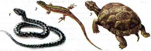
Rice. 141. Different kinds reptiles: 1 - common viper; 2 - nimble lizard; 3 - steppe turtle
Due to the terrestrial lifestyle and the transition to exclusively pulmonary respiration, the body of reptiles is covered with horny scales and lacks glands.
The skeleton of reptiles, to a greater extent than of amphibians, is adapted to life on land (Fig. 142, B). The skull has one protrusion - the condyle, with which the back of the skull is attached to the spine. This makes the head very mobile. The spine of a lizard is divided into five sections: cervical, thoracic, lumbar, sacral and caudal. V cervical spine- 7-10 movable vertebrae. The first two stand out - the atlas and the epistrophy. Their articulation increases the mobility of the head.

Rice. 142. Lizard body structure: A - external structure: 1 - head; 2 - torso; 3 - tail; 4 - front limbs; 5 - hind limbs; B - skeleton: 1 - spine; 2 - skull; 3 - the belt of the forelimbs; 4 - the belt of the hind limbs; 5 - shoulder; 6 - forearm; 7 - brush; 8 - thigh; 9 - shin; 10 - foot
The ribs are attached to the trunk vertebrae (16-25). The front ribs connecting to the sternum form the rib cage. It protects the organs in the chest cavity (esophagus, trachea, heart, lungs) from damage and participates in the respiratory mechanism: it expands when inhaling and falls off when exhaling. The ribs in snakes are attached to the vertebrae of the entire trunk of the spine and do not connect to the sternum: snakes have no chest.
The hind limb is attached to the sacral vertebrae (there are two of them) in the lizard. The skeleton of the girdles and free limbs retains the general structural scheme characteristic of all terrestrial vertebrates. The limbs of the lizards are widely spaced apart, but there are also legless lizards among the lizards. Snakes also have no legs. In these cases, reptiles move with powerful muscles attached to the spine and ribs, the ends of which protrude through the skin and cling to uneven ground.
Exercises on the covered material
- Explain the origin of the class name Reptiles. Give examples to support this name.
- What adaptations of the external structure provide the terrestrial way of life for reptiles?
- What structural features of the skeleton of reptiles are associated with their life on land?
- What are the vital processes of reptiles that provide them with life on land?
-
 April 17, 2015True Tales of the Brothers Grimm
 -
 April 17, 2015The best works of Tolstoy for children
 -
 April 17, 2015Leo Tolstoy all the best tales and stories


